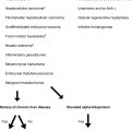
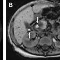
The differential diagnosis of renal masses in pediatric patients includes benign and malignant tumors, as well as nonneoplastic mass-like lesions mimicking tumors. Although the spectrum of renal masses in children has some overlap with that of adults, it is important to understand the renal pathologic processes specific to the pediatric population, as well as their characteristic imaging appearances and clinical presentations. This article reviews benign and malignant renal masses in children, with an emphasis on magnetic resonance imaging and clinical features that are specific to each lesion type.
Key points
- •
Children younger than 7 to 9 years may have difficulty undergoing renal magnetic resonance (MR) imaging because of anxiety and/or inability to lie still. Distraction techniques and sedation/general anesthesia are commonly used methods for helping young children cooperate with renal MR imaging.
- •
At present, renal MR imaging is performed at field strength of either 1.5 or 3-T. While the current trend is toward imaging at higher field strength, it is important for pediatric radiologists to understand the advantages and disadvantages of MR imaging at higher field strength as it applies to renal imaging.
- •
Angiomyolipoma is a benign renal mass that can often be diagnosed conclusively on MR imaging, owing to the presence of internal adipose tissue components. Fat-poor angiomyolipomas, however, pose a diagnostic dilemma and can be difficult to distinguish from malignancy.
- •
For malignant renal masses, MR imaging is very useful for detecting synchronous renal masses, abdominal metastasis, and extrarenal/vascular tumor invasion, all of which have important therapeutic implications.
- •
Patient age, clinical symptoms, and syndromic associations are important ancillary data for narrowing down the differential diagnosis of a pediatric renal mass.
Stay updated, free articles. Join our Telegram channel

Full access? Get Clinical Tree


