MALIGNANT AND OTHER AGGRESSIVE TUMORS OF VASCULAR ORIGIN
KEY POINTS
- Malignant vascular neoplasms are uncommon and can mimic traditionally hypervascular and more common head and neck region masses such as paragangliomas.
- Proliferative hemangioma can mimic “malignant” behavior when it grows particularly rapidly.
- Hypervascularity may be a reactive change and associated with other benign processes.
ANGIOSARCOMA
Clinical Perspective and Pathology
Angiosarcomas are rare sarcomas of blood vessel or lymphatic vessel origin.1 Those most commonly encountered arise in the head and neck region mainly from the scalp and face.2 These very aggressive malignancies grow in a highly infiltrative manner, destroy bone, and metastasize early to regional nodes and distant sites (Fig. 25.1).
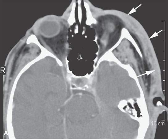
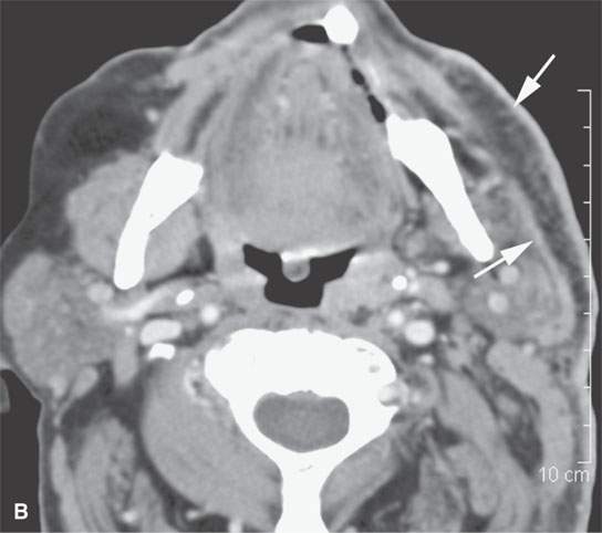
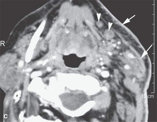
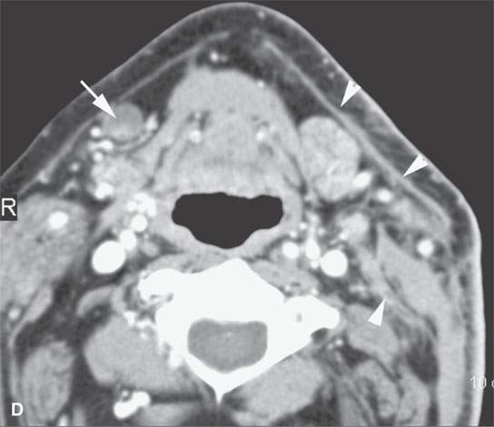
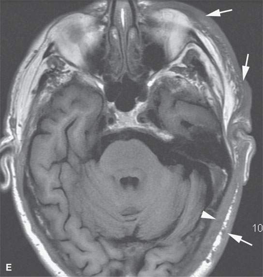
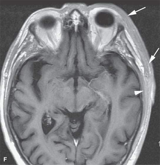
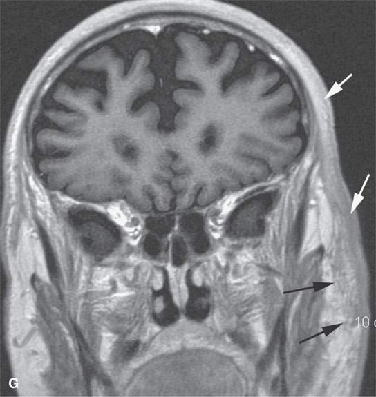
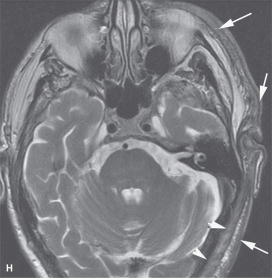
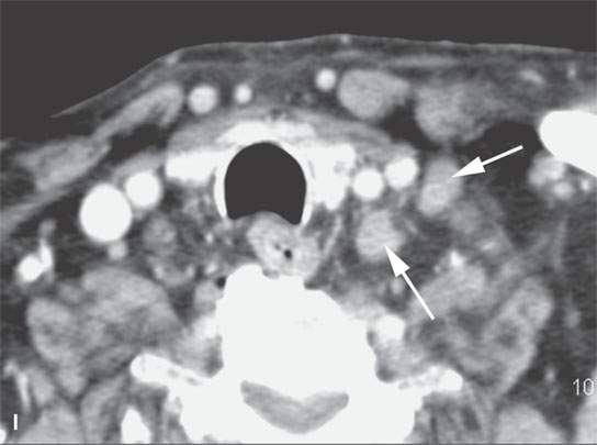
FIGURE 25.1. Computed tomography (CT) (A–D, I) and magnetic resonance imaging (E–H) in a patient with angiosarcoma of the skin. A: Extensive thickening and enhancement of the skin (arrows). B: Extensive thickening and enhancement of the skin, at least edema, involving the subcutaneous fat and superficial musculoaponeurotic system (SMAS) (arrows). C: Extensive thickening and enhancement of the skin and SMAS (arrows) with metastatic deposits in level 1B nodes (arrowheads). D: Very extensive thickening and enhancement of the skin, SMAS, and deeper fascia planes likely due to lymphatic engorgement and diffuse tumor seeding (arrowheads) with metastatic deposits in a contralateral level 1B node (arrow). E: Non–contrast-enhanced T1-weighted (T1W) image with extensive skin (arrows) and deeper scalp muscle thickening (arrowhead). F: Contrast-enhanced T1W image with extensive skin (arrows) and deeper scalp muscle thickening and enhancement (arrowhead). G: Contrast-enhanced T1W image with extensive skin and deeper scalp muscle thickening and enhancement (white arrows) and nodular enhancement in the subcutaneous and deeper fat (black arrows). H: T2-weighted image with extensive skin (arrows) and deeper scalp muscle thickening (arrowheads). I: Contrast-enhanced CT showing metastatic nodes (arrows) in levels 5B and 6.
Angiosarcomas occur mainly in patients older than 65 years of age but can be seen at any age down to 2 years.3,4 Males are more likely to be affected.4 Prognosis is poor.5 Surgery is the preferred method of treatment. Radiation is considered palliative or adjunctive.1,5
Imaging Appearance
Angiosarcomas enhance brightly on contrast-enhanced computed tomography (CECT) and contrast-enhanced magnetic resonance (CEMR)5 (Fig. 25.1).5 On T2-weighted magnetic resonance (MR), the mass is very bright and has no discernible border with surrounding edematous tissue. The superficial type may be hard to appreciate on any imaging study compared to physical examination. All head and neck nodal groups should be included as discussed in conjunction with skin cancers (Fig. 25.1I) (Chapter 24).
HEMANGIOPERICYTOMA
Clinical Perspective and Pathology
Hemangiopericytoma is a neoplasm composed of capillaries surrounded by an accumulation of spindle or round cells.6,7 The round cells come from the pericyte of Zimmerman.7,8 It has been suggested that the pericyte controls the caliber of the vessel. The hallmark of the tumor is its variability in appearance, growth, and biologic behavior (Figs. 25.2–25.4). Depending on their degree of histologic aggressiveness, these lesions tend to metastasize very frequently.
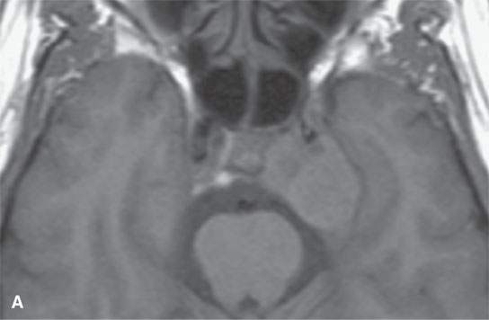
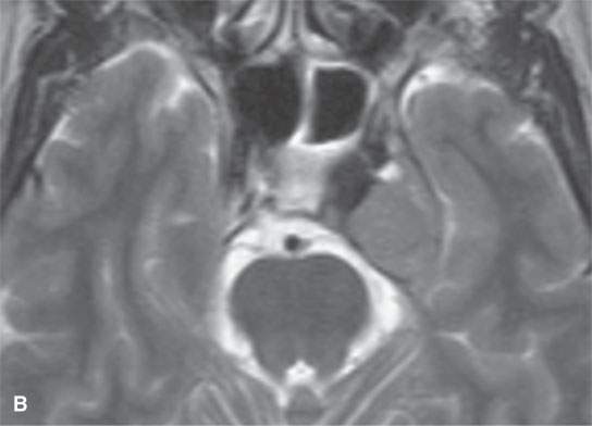
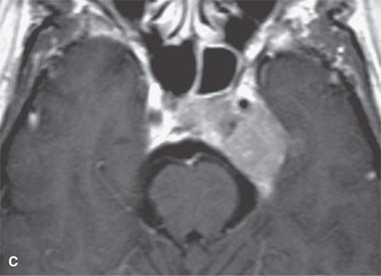
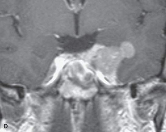
FIGURE 25.2. Contrast-enhanced magnetic resonance of a relatively benign-appearing hemangiopericytoma, with the only feature somewhat atypical for a meningioma being the nodule of exophytic growth. This was thought to be a meningioma prior to biopsy. A: Non–contrast-enhanced T1-weighted (T1W) image. B: T2-weighted image. C: Contrast-enhanced T1W image. D: Coronal contrast-enhanced T1W image showing an exophytic nodule.
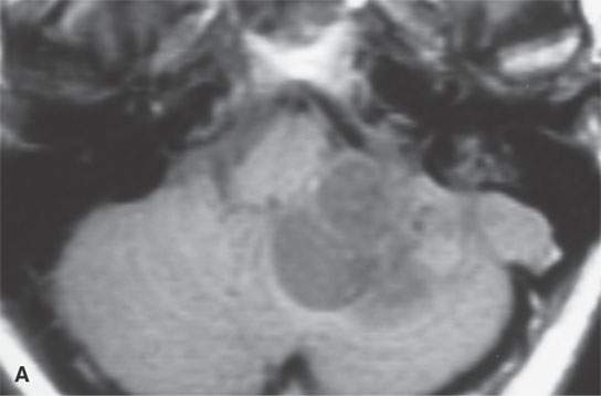
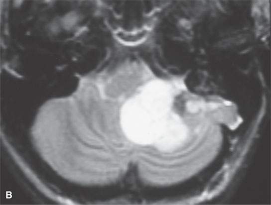
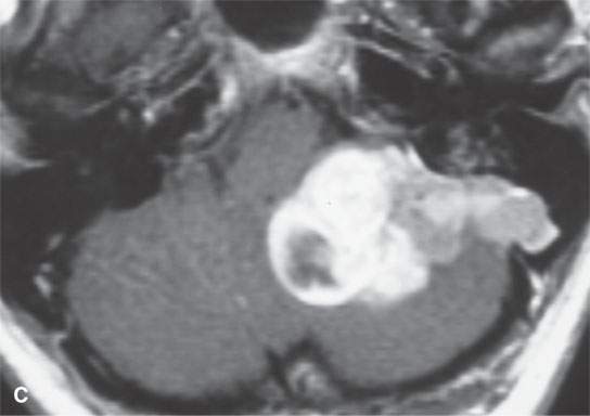
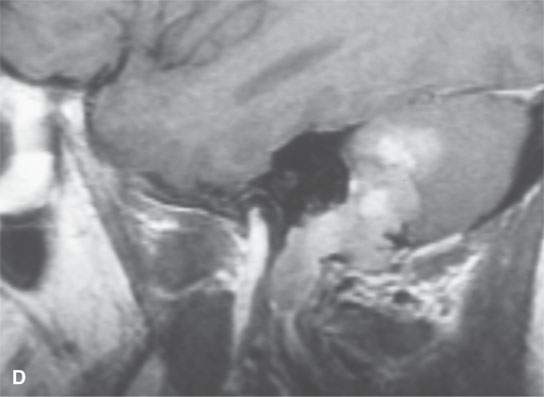
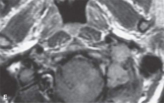
FIGURE 25.3. Contrast-enhanced magnetic resonance of a relatively low-grade hemangiopericytoma invading and growing within the sigmoid sinus and along or within jugular vein. A: Non–contrast-enhanced T1-weighted (T1W) image. B: T2-weighted image. C: Contrast-enhanced T1W image. D, E: Contrast-enhanced T1W images showing the transcranial extension along the jugular vein and growth into the soft tissues below the skull base.
Related posts:
 Preseptal Compartment: Acute and Chronic Infections and Noninfectious Inflammatory Conditions
 Necrotizing (“Malignant”) Otitis Externa
 Cervicothoracic Junction: Brachial Plexus Trauma
 Oropharynx: Introduction
 Branchial Apparatus Developmental Anomalies
 Oral Cavity and Floor of the Mouth: Malignant Tumors
Stay updated, free articles. Join our Telegram channel

Full access? Get Clinical Tree


