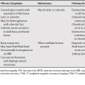
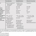
123 Magnetic resonance imaging (MRI) may be helpful in differentiating benign from malignant soft tissue lesions, although there is conflicting evidence in the literature. In published prospective studies, the sensitivity ranges from 78 to 100%, and the specificity from 17 to 89%.1 Specific diagnoses are sometimes possible based on signal intensity (lipoma, fibrous lesions), or signal intensity and lack of enhancement (cyst, ganglion). Vascular lesions such as soft tissue hemangiomas (lobulation, septation, and low-signal intensity dots)2 can often be definitively diagnosed. Note that, unlike malignant bone tumors, malignant soft tissue masses may have well-defined margins; thus, lesion margins are usually not a helpful differentiating factor.3 In particular, synovial sarcomas are often small with well-defined margins.4
Malignant versus Benign Soft Tissue Lesions
Stay updated, free articles. Join our Telegram channel

Full access? Get Clinical Tree


