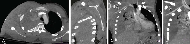
Management of Extremity Vascular Trauma

Chapter 39


Management of Extremity Vascular Trauma


Sean R. Dariushnia and Curtis A. Lewis


Vascular injury has two main consequences—hemorrhage and ischemia. Or, in the words of an anonymous Czech surgeon, “Bloody vascular trauma—it’s either bleeding too much or it’s not bleeding enough.”1 Unrecognized and uncontrolled hemorrhage can rapidly lead to a trauma patient’s demise. Unrecognized and untreated ischemia can lead to limb loss, stroke, bowel necrosis, and multiple organ failure. The aim of this chapter is to highlight the fundamentals of peripheral vascular trauma and provide an approach to the diagnosis and management of vascular injury.


Arterial and venous structures are most commonly injured by penetrating trauma, with a much higher incidence in gunshot wounds than for stab injury. Blunt trauma also carries a significant vascular injury rate, and iatrogenic vascular injuries are increasing as radiologic and minimal-access procedures become more commonplace.


Hemorrhage is the prime consequence of vascular injury. Bleeding may be obvious, with visible arterial hemorrhage, or it may be concealed. Classically, concealed arterial hemorrhage may be in the chest, abdomen, and pelvis. Hemorrhage may also be concealed in the soft tissues of the buttock and thigh, and blood from facial fractures may be swallowed and go unnoticed.


Ischemia results from acute interruption of blood flow to a limb or organ. When oxygen supply is inadequate to meet demand, anaerobic metabolism takes over, producing lactic acidosis and activating cellular and humoral inflammatory pathways. If the arterial supply is not reestablished in time, cell death occurs. Skeletal muscle can be rendered ischemic for 3 to 6 hours and still recover function. Peripheral nerves are more sensitive to ischemia, and prolonged neurologic deficit may result from relatively short periods of tissue ischemia.


If arterial supply is restored to ischemic tissue, the sudden release of inflammatory mediators, lactic acid, potassium, and other intracellular material into the circulation can cause profound myocardial depression and generalized vasodilatation and may initiate a systemic inflammatory response.


Vascular trauma can result in serious and potentially fatal consequences. Percutaneous intervention can provide immediate control of hemorrhage and in many cases can be lifesaving.


In addition, definitive long-lasting treatments can be obtained using the percutaneous approach when patients are appropriately selected. In view of the rising number of cases of vascular trauma seen at trauma centers, endovascular therapy has an increasing role in the treatment of traumatic vascular injury.


Indications


Patterns of Vascular Injury


Laceration with either complete or incomplete vessel transection is the most common form of vascular injury. Hemorrhage tends to be more severe in partially transected vessels because complete transection results in retraction and vasoconstriction of the vessel, limiting or even preventing arterial hemorrhage.


Blunt trauma injures vessels by crushing, distraction, or shearing. This results in contusion to the vessel, which may extend for some distance along its length. An intimal flap may be formed that will lead to thrombosis or dissection and subsequent rupture. Thrombosis may propagate for some distance down the vessel, or there may be embolization to produce effects more distally.


Arterial hemorrhage may continue within a contained hematoma, leading to a pulsatile mass, a pseudoaneurysm. Commonly, distal flow is preserved with false aneurysm formation, and diagnosis may be difficult. Aneurysms are at risk of rupture if undiagnosed, and often present late after the initial injury is forgotten.


If there is injury to an adjacent vein as well as to the artery, an arteriovenous fistula (AVF) can form, which may subsequently lead to rupture or cardiovascular compromise. AVFs may present some time after the initial injury.


Spasm as a unique entity is never the result of trauma and should not be assumed to be the cause of limb ischemia.


Technique


The diagnosis of significant vascular injury rests almost entirely on the physical examination. An absence of hard signs of vascular injury does not exclude the presence of vascular trauma. In contrast, the presence of hard signs mandates immediate action.


Diagnostic Tests


Doppler Ultrasonography


The diagnosis of a significant (i.e., requiring intervention) vascular injury relates to the presence or absence of a palpable pulse. Presence of a Doppler signal in a pulseless limb only gives a false sense of security and does not imply a less severe or less urgent injury pattern. A diminished but palpable pulse is a soft sign of vascular injury.


Similarly, a reduction in the ankle-brachial pressure index in the presence of a palpable pulse does indicate the presence of a vascular injury requiring intervention. A differential in peak systolic cuff pressures between the traumatized and normal extremity predicts a significant (requiring repair) vascular injury at angiography. This was clearly demonstrated in 1993 by Schwartz and Weaver, who entered over 500 consecutive patients with isolated upper or lower extremity penetrating injury into a prospective study designed to refine the indications for diagnostic arteriography. Fourteen major injuries and 10 nonocclusive injuries were identified. Only pulse deficit (P < .01) and an ankle-brachial or wrist-brachial index less than 1.00 in the injured extremity (P < .03) were found to be significant predictors of an arterial injury.2 Doppler sonography alone, therefore, adds little to careful clinical examination.


Computed Tomographic Angiography


CT scanners are present in nearly every hospital. In trauma centers, 16- and 64-MDCT technology is commonplace and allows for fast image acquisition of the entire body (seconds). Routinely, if the patient presents with polytrauma and is hemodynamically stable, a head, chest, and abdomen/pelvis CT (PAN-SCAN) can be obtained. With multidetector scanners, an extremity computed tomographic angiography (CTA) examination can easily be added to the PAN-SCAN with the same contrast injection, without significantly adding to image acquisition time.3 Axial images can be viewed immediately after image acquisition and most injuries identified. Postprocessing three-dimensional (3D) reconstructions can provide an easily understandable format obtainable within minutes of the scan and can be of benefit to both the radiologist and nonradiologist clinician, especially the vascular or trauma surgeon (Fig. 39-1).


Stay updated, free articles. Join our Telegram channel

Dec 23, 2015 | Posted by in INTERVENTIONAL RADIOLOGY | Comments Off on Management of Extremity Vascular Trauma

Full access? Get Clinical Tree

Get Clinical Tree app for offline access