Management of Failing Hemodialysis Access


Management of Failing Hemodialysis Access


Charles T. Burke


The natural history of a vascular access graft or fistula is progressive development of neointimal hyperplastic stenoses that reduces blood flow and compromises performance of the vascular access. If left untreated, these lesions will eventually lead to thrombosis of the vascular access. Early detection and treatment of hemodynamically significant stenosis is a primary tenet of a vascular access management program. Angioplasty remains the most important technique for treating neointimal hyperplastic stenoses associated with hemodialysis grafts and fistulas.


Indications


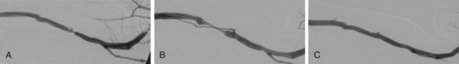
The criteria that define failing vascular access and the indications for percutaneous or surgical repair are described in the National Kidney Foundation (NKF) Clinical Practice Guidelines for Vascular Access.1 These criteria and indications have been incorporated into the Society of Interventional Radiology (SIR) quality improvement guidelines, the American College of Radiology (ACR)-SIR practice guidelines, and the SIR document on standards of reporting.13


Primary indications for percutaneous intervention of either polytetrafluoroethylene (PTFE) grafts or arteriovenous fistulas (AVFs) include (1) documentation of a clinical or hemodynamic abnormality and (2) a stenosis causing over 50% reduction in luminal diameter. Intervention is not indicated unless the clinical and angiographic findings are both abnormal and correlative. Clinical and hemodynamic abnormalities indicative of significant stenosis include elevated venous pressure, decreased intra-access blood flow, prolonged bleeding after needle removal, and arm swelling.


Several clinical studies have reported that periodic assessment of intra-access blood flow may be the most reliable method for early detection of a developing stenosis.46 Intra-access blood flow is measured during hemodialysis, and if intra-access blood flow is less than 600 mL/min, a fistulogram is recommended to evaluate the entire vascular access circuit.1


Over time, there may be degeneration of graft material, resulting in overlying pseudoaneurysms. This is often due to fragmentation of the graft material from repeated cannulation and increased intragraft pressure from venous outflow stenosis.7 Treatment may be required when the pseudoaneurysm interferes with graft access or there are signs of impending rupture. Indicators for possible graft rupture include skin breakdown over the pseudoaneurysm or rapid pseudoaneurysm expansion. In severe cases or in the setting of graft infection, pseudoaneurysms are treated surgically. In patients with limited surgical options and in whom the anatomy is suitable, endovascular management may be considered.


Early evaluation and intervention may be useful for salvage of nonmaturing fistulas.8 Recent reports demonstrate that serial dilation of small-diameter veins (balloon maturation) may expedite the maturation process and provide functionality to fistulas that may otherwise have failed.911 Although this indication for percutaneous intervention is not yet fully defined, AVFs that have failed to mature within 3 months of creation should undergo fistulography to determine whether angioplasty or surgical revision would be beneficial. The primary reasons for a fistula that fails to mature include vascular stenosis (outflow vein or arteriovenous anastomosis), competitive outflow veins, or a deep outflow vein that is nonpalpable. Management of vascular stenoses continues to be balloon angioplasty. Small competitive outflow veins can be managed by surgical ligation or endovascular embolization. Deep nonpalpable outflow veins require surgical revision.


Use of stents or stent-grafts for management of failing hemodialysis grafts and fistulas remains controversial. Although angioplasty remains the primary technique for treating neointimal hyperplastic stenosis, stent placement may be indicated in specific situations. The Kidney Disease Outcomes Quality Initiative (KDOQI) guidelines allow for stent use to treat central venous stenoses when there is (1) acute elastic recoil of the vein and over 50% residual stenosis after angioplasty and (2) stenosis that recurs within 3 months after previously successful angioplasty.1 When treating an aggressive recurring stenosis in a large central vein, a vascular stent may provide better results than angioplasty alone. Another acceptable use of stents is for acute repair of angioplasty-induced venous rupture. Although minor venous injuries can be effectively managed by prolonged inflation of an angioplasty balloon, more substantial injuries with continuing perivascular hemorrhage may require a stent or stent-graft to provide a more durable repair.12,13


Contraindications


The only absolute contraindication to percutaneous intervention is an infected vascular access. Manipulation of an infected graft or fistula may cause intravascular dissemination of microorganisms and thereby elicit acute sepsis or long-term infectious complications such as endocarditis or osteomyelitis. Superficial cellulitis may resolve with appropriate antibiotic treatment, but more extensive infection of a graft or fistula often requires surgical management.


Another important contraindication to percutaneous intervention is allergy to a contrast agent. Patients with a known or suspected allergy should be pretreated according to standardized protocols before fistulography or any interventional procedure requiring use of intravascular contrast material. Alternatively, a noniodinated contrast agent (e.g., CO2) may be used.14


Relative contraindications include intervention on a newly placed graft or fistula; dilation of a new (<30 days) anastomosis may disrupt its integrity. Significant anastomotic stenosis within 30 days after creation of the vascular access suggests a technical or anatomic problem that often requires surgical revision to achieve optimal long-term patency.


Long-segment venous stenoses and occlusions are also relative contraindications to percutaneous management. Compared with treatment of short (<3 cm) lesions, angioplasty of long (>7 cm) venous stenoses and occlusions often results in suboptimal immediate success and reduced long-term patency. Although a stent or stent-graft can be inserted to improve postangioplasty luminal diameter, the long-term durability of such interventions remains questionable. When confronted with long lesions or multiple segments of venous stenosis, the appropriate management decision should include both percutaneous and surgical options.


Equipment


Though once the domain of hospital-based practices, an increasing number of vascular access–related procedures are being performed in outpatient clinics and freestanding ambulatory surgery centers. Regardless of the setting, the equipment required includes (1) a dedicated procedural room, (2) a digital subtraction angiography system, (3) physiologic monitoring and resuscitation equipment, and (4) procedural supplies such as angiographic catheters, guidewires, and needles.


The extent and type of angiography supplies are dependent on physician preference and the variety of procedures performed. A selection of multipurpose angiographic catheters, guidewires, and vascular sheaths is necessary for basic interventional procedures. High-pressure angioplasty balloons with rated burst pressures of greater than 15 atm are often required for effective treatment of neointimal hyperplastic stenoses. Balloon diameters from 5 to 14 mm are required to treat both peripheral and central stenoses. For very fibrotic and resistant stenoses, a peripheral cutting balloon may be useful.


Although stents and stent-grafts are not frequently used for vascular access–related problems, it is prudent to have several readily available if needed to repair an acute procedure-related complication. Both balloon-expandable and self-expandable stents should be available. Each type has advantages and disadvantages, which should be taken into account when determining appropriate situations for their use.


More advanced procedures may require additional supplies such as microcatheters and microguidewires, embolic agents such as coils, and snares.


Technique


Anatomy and Approach


For the relevant venous anatomy, please refer to Chapters 30 and 82.


Physical examination of the vascular access is a reliable method for determining the approximate location of stenoses before fistulography. Information gained from the physical examination should be used to plan an appropriate site for entry into the vascular access. In some instances, particularly with the complicated anatomy of an AVF, a preprocedural ultrasound examination can be useful to locate stenoses and plan the optimal entry site for subsequent interventions.


Assessment of the entire vascular access circuit should be performed before embarking on a reparative procedure. The appropriateness of a percutaneous intervention depends on numerous factors, including the location and length of the lesion, number of lesions, and patency of contiguous and adjacent veins. The complex anatomy of AVFs can present a challenge to novice interventionalists. An understanding of standard fistula configurations is essential for correct interpretation of angiographic images. Evaluation of a nonmaturing fistula can be particularly difficult because of the complexity of the anatomy, the small caliber of the veins, and the multiplicity of problems that can be found during fistulography. It is often useful to review the patient’s surgical vascular access records when confronted with unusual or complex vascular anatomy.


Technical Aspects


Fistulography


A diagnostic fistulogram can be performed by inserting a 21-gauge butterfly needle or a Venflon access cannula into the hemodialysis graft or fistula and injecting small aliquots (<5 mL) of radiographic contrast material to opacify the different segments of the vascular access circuit. Some practitioners use a micropuncture vascular access set and insert the 5F dilator into the vascular access for injection of contrast material. The fistulogram is performed to evaluate the hemodialysis graft or fistula and the entire vascular access circuit from the arterial anastomosis to the superior vena cava. For nonmaturing fistulae, ultrasound is often required to identify the primary outflow vein. Occasionally, antegrade micropuncture of the brachial artery is necessary to outline the vascular anatomy. Endovascular interventions would be performed via a subsequent outflow vein access.


The majority of patients referred for fistulography will have at least one significant lesion requiring percutaneous intervention. Ideally the initial entry site into the vascular access should be chosen to provide a favorable route for subsequent interventional procedures. Once identified, the character, extent, and hemodynamic significance of each lesion should be assessed before proceeding with a percutaneous intervention. Multiple angiographic images obtained in different imaging planes are often necessary to thoroughly evaluate a stenosis before proceeding with percutaneous treatment. When measured on a two-dimensional angiographic image, a 50% luminal stenosis corresponds to a 75% to 80% decrease in cross-sectional area and is considered hemodynamically significant.15 The fistulogram may also demonstrate the presence of venous collateral channels adjacent to a stenosis, a finding indicative of significant obstruction.


The hemodynamic significance of a stenosis can be difficult to determine with angiographic imaging alone.16 Quantitative measurements can be performed during the procedure, both before and after intervention, to ascertain a lesion’s hemodynamic significance. Pullback pressure measurements can be used to measure the transstenotic pressure gradient: a mean pressure gradient above 10 mmHg represents hemodynamically significant stenosis.17 Intraprocedural measurement of intra-access blood flow using specialized thermodilution catheters can also provide quantitative hemodynamic information.18 This technique is particularly useful for assessing the functional significance of complicated or multifocal stenoses.


When performing fistulography, it is important to distinguish between fixed stenoses and venous spasm. Venous spasm typically appears as a long, smoothly tapered stenosis in a native vein, often at or near the point of access. Administration of a vasodilator drug (e.g., nitroglycerin) directly into the abnormal venous segment may help differentiate venous spasm from fixed stenosis. Venous spasm will often resolve after vasodilator administration, whereas a true stenosis will persist (Fig. 115-1).


Angioplasty


Percutaneous transluminal angioplasty (PTA) is the most commonly performed percutaneous procedure for treating neointimal hyperplastic stenosis associated with hemodialysis grafts and fistulas. The operator should be prepared to perform angioplasty after completion of the diagnostic fistulogram when an appropriate lesion is identified.


As described earlier, the initial entry site into the vascular access should be planned to allow easy access to stenotic or occluded segments of the vascular access circuit. This may require obtaining new vascular access at a new site or in the opposite direction. Though not mandatory, a vascular sheath can prove advantageous for injection of contrast material and emergency maneuvers in the event of an angioplasty-induced complication. Anticoagulants are not uniformly administered for routine angioplasty procedures.

Stay updated, free articles. Join our Telegram channel

Dec 23, 2015 | Posted by in INTERVENTIONAL RADIOLOGY | Comments Off on Management of Failing Hemodialysis Access

Full access? Get Clinical Tree

Get Clinical Tree app for offline access