Presentation and Presenting Images
A 40- year-old female presents for screening mammography.
28.2 Key Images
28.2.1 Breast Tissue Density
There are scattered areas of fibroglandular density.
28.2.2 Imaging Findings
In the upper outer quadrant of the right breast at the 10 o’clock location in the middle depth, there is an oval high-density mass. The digital breast tomosynthesis (DBT) images ( ▶ Fig. 28.3 and ▶ Fig. 28.4) suggest that the margins of the mass are mostly circumscribed. The left breast imaging (not shown) was normal.
28.3 BI-RADS Classification and Action
Category 0: Mammography: Incomplete. Need additional imaging evaluation and/or prior mammograms for comparison.
28.4 Diagnostic Images
( ▶ Fig. 28.5, ▶ Fig. 28.6, ▶ Fig. 28.7, ▶ Fig. 28.8, ▶ Fig. 28.9)
28.4.1 Imaging Findings
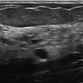
The diagnostic imaging demonstrates a 1.2-cm oval mass with obscured and circumscribed margins in the right breast in the middle depth. The targeted ultrasound demonstrates a 1.2 × 0.9 × 1.2 cm anechoic oval mass, 7 cm from the nipple, with circumscribed margins, mild posterior acoustic enhancement, and no internal or surrounding vascularity.
28.5 BI-RADS Classification and Action
Category 2: Benign
28.6 Differential Diagnosis
Simple cyst: This mass on mammography is circumscribed; however, the ultrasound confirms that this is a fluid-containing mass and its imaging features also suggest this is a simple cyst.
Complicated cyst: Complicated cysts typically have low-level internal echoes and have a look similar to solid masses. This mass has no internal echoes. Sometimes cysts deep in the breast tissue can be difficult to visualize with ultrasound. Thus, it is important that the gain and frequency of the ultrasound machine is properly adjusted to allow for the distinction between cysts and solid masses to be perceived on ultrasound imaging.
Fibroadenoma: This mass appears fluid-filled thus eliminating fibroadenoma as a diagnosis. On baseline mammograms of young women, the most common masses will be cysts or fibroadenomas.
28.7 Essential Facts
Recall rates are decreased with DBT compared to full-field digital mammography (FFDM).
Durand and colleagues (2015) found that baseline recall rates for DBT and FFDM were significantly reduced compared to FFDM alone (20.8% compared with 33.1%, respectively).
Masses seen on a baseline DBT compared with a baseline FFDM had no significant difference in recall rates.
There is a significant reduction in recalls for asymmetries seen on baseline FFDM and DBT mammograms compared to asymmetries seen on baseline FFDM alone.
28.8 Management and Digital Breast Tomosynthesis Principles
The unmasking effect of DBT and the ability to present the slices within a three-dimensional (3D) volume allow for better characterization of the tissue composition and lesions seen within the tissue.
DBT is superior to FFDM in characterizing the margins of a mass.
There is not enough experience with DBT to dismiss masses that are completely circumscribed on DBT as benign (BIRADS 2) and in need of no further evaluation.
28.9 Further Reading
[1] Durand MA, Haas BM, Yao X, et al. Early clinical experience with digital breast tomosynthesis for screening mammography. Radiology. 2015; 274(1): 85‐92 PubMed
[2] Helvie MA. Digital mammography imaging: breast tomosynthesis and advanced applications. Radiol Clin North Am. 2010; 48(5): 917‐929 PubMed
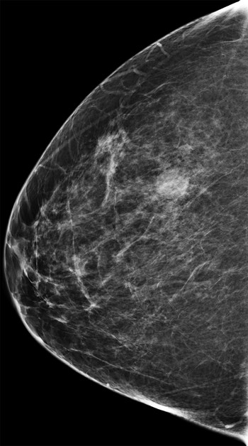
Fig. 28.1 Right craniocaudal (RCC) mammogram.
Related posts:


Stay updated, free articles. Join our Telegram channel

Full access? Get Clinical Tree


