Presentation and Presenting Images
A 43-year-old female presents for baseline screening mammography.
32.2 Key Images
32.2.1 Breast Tissue Density
There are scattered areas of fibroglandular density.
32.2.2 Imaging Findings
Central to the nipple in the anterior depth, there is an oval mass. It appears circumscribed on the craniocaudal (CC) view ( ▶ Fig. 32.1) and partially obscured on the mediolateral (MLO) view ( ▶ Fig. 32.2). The digital breast tomosynthesis (DBT) images confirm that this mass is circumscribed ( ▶ Fig. 32.3 and ▶ Fig. 32.4).
32.3 BI-RADS Classification and Action
Category 0: Mammography: Incomplete. Need additional imaging evaluation and/or prior mammograms for comparison.
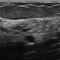
32.4 Diagnostic Images
( ▶ Fig. 32.5, ▶ Fig. 32.6, ▶ Fig. 32.7, ▶ Fig. 32.8)
32.4.1 Imaging Findings
The diagnostic imaging demonstrates the oval 16 × 11 × 10 mm mass with circumscribed margins at the 9 o’clock location in the anterior depth ( ▶ Fig. 32.5, ▶ Fig. 32.6, and ▶ Fig. 32.7). The corresponding ultrasound reveals a hypoechoic oval mass that measures 15 × 6 × 15 mm ( ▶ Fig. 32.8). There is no increase in vascularity.
32.5 BI-RADS Classification and Action
Category 3: Probably benign
32.6 Differential Diagnosis
Fibroadenoma: This mass is oval and circumscribed and has benign features. With this being a baseline mammogram, it is reasonable to follow benign-appearing masses or offer a biopsy. The patient elected for observation. Her follow-up evaluations at 6 and 12 months revealed no change.
Complicated cyst: The homogeneous low-level internal echoes that can be present in a complicated cyst can be difficult to distinguish from a solid mass. If there were internal vascularity present, this would exclude a complicated cyst.
Phyllodes tumor: These tumors are not as common as fibroadenomas, but they can often mimic each other. These typically grow over time and would require intervention if growth were seen during a period of observation.
32.7 Essential Facts
Fibroadenomas are fibroepithelial tumors with stromal and epithelial elements.
Fibroadenomas are the most common solid lesions of the breast.
Most fibroadenomas are unilateral, but up to 20% of patients will have bilateral lesions.
Recall rates tend to be higher in women receiving a baseline mammogram.
Due to the increase in specificity without a decrease in sensitivity for patients who have full-field digital mammography (FFDM) with digital breast tomosynthesis (DBT) when receiving a baseline mammogram, there has been a decrease in the recall rate for these patients.
A reduction in recall rates for DBT were seen irrespective of breast tissue density.
32.8 Management and Digital Breast Tomosynthesis Principles
DBT can detect many benign lesions (fibroadenomas and cysts). It is uncertain if these lesions can be classified as benign based on their round or oval shape with circumscribed margins. Large-scale studies are needed to determine if these lesions can be dismissed on DBT and, if so, what the strict criteria are to do so.
A mass with classic benign features on a baseline mammogran can be assigned a BI-RADS 3 category and followed. The typical time course for following is 2 years of stability, with imaging at 6 months, 12 months, and 24 months. However, if this mass is a new finding on a mammogram (one with comparisons), it should be assigned a BI-RADS 4a category and biopsy should be recommended.
32.9 Further Reading
[1] Baker JA, Lo JY. Breast tomosynthesis: state-of-the-art and review of the literature. Acad Radiol. 2011; 18(10): 1298‐1310 PubMed
[2] Sumkin JH, Ganott MA, Chough DM, et al. Recall Rate Reduction with Tomosynthesis During Baseline Screening Examinations: An Assessment From a Prospective Trial. Acad Radiol. 2015; 22(12): 1477‐1482 PubMed

Fig. 32.1 Right craniocaudal (RCC) mammogram.
Related posts:


Stay updated, free articles. Join our Telegram channel

Full access? Get Clinical Tree


