Presentation and Presenting Images
A 49-year-old female who has a sister with breast cancer presents for screening mammography.
25.2 Key Images
( ▶ Fig. 25.3, ▶ Fig. 25.4, ▶ Fig. 25.5, ▶ Fig. 25.6)
25.2.1 Breast Tissue Density
There are scattered areas of fibroglandular density.
25.2.2 Imaging Findings
In the upper outer quadrant of the right breast at the 9 o’clock location in the posterior depth, there is a possible focal asymmetry. This is best seen on the digital breast tomosynthesis (DBT) images, especially the mediolateral oblique (MLO) DBT images. There does not initially appear to be a correlate on the corresponding MLO mammogram. The DBT images reveal a mass with indistinct margins at the 9 o’clock location that appears larger on the craniocaudal (CC) DBT images than on the MLO DBT images.
25.3 BI-RADS Classification and Action
Category 0: Mammography: Incomplete. Need additional imaging evaluation and/or prior mammograms for comparison.
25.4 Diagnostic Images
( ▶ Fig. 25.7, ▶ Fig. 25.8, ▶ Fig. 25.9)
25.4.1 Imaging Findings
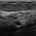
The diagnostic imaging demonstrates a persistent mass on the CC spot-compression mammogram. The ML mammogram suggests a mass at the 9 o’clock location but the density of the breast interferes with further assessment. An ultrasound was performed because the DBT images were convincing that there was a mass present and the margins were indistinct. The ultrasound evaluation revealed an 8-mm anechoic mass with circumscribed margins and posterior acoustic enhancement surrounded by dense breast tissue.
25.5 BI-RADS Classification and Action
Category 2: Benign
25.6 Differential Diagnosis
Simple cyst: The adjacent dense tissue on conventional mammography and DBT made this mass appear bigger than it is. Ultrasound reveals an anechoic mass with circumscribed margins mostly surrounded by dense tissue.
Solid mass: Neither conventional mammography nor DBT can tell a fluid-filled (cystic) mass from a solid mass. The ultrasound appearance is highly suggestive of a fluid-filled structure.
Normal fibroglandular tissue: Conventional mammography was less convincing of the presence of any finding. The ability of DBT to decrease the effects of overlapping tissue can lead to the identification of both malignant and benign findings.
25.7 Essential Facts
Lesion margins become more apparent with DBT.
Oval or round masses with gently lobulated margins will be unmasked with DBT when previously concealed on conventional breast imaging. Further studies will be needed to determine whether these masses can be safely followed or ignored.
Summation artifacts are a known cause of false-positives on screening mammography. DBT can help to minimize this contribution to false-positives by demonstrating that the finding is due to overlapping fibroglandular tissue.
25.8 Management and Digital Breast Tomosynthesis Principles
There is a learning curve with DBT and readers must reset their threshold for calling back findings on DBT for further evaluation.
DBT will unmask small circumscribed masses, which are probably cysts or intramammary lymph nodes, that were previously obscured.
It is uncertain how the unmasking effect of presumably benign findings will affect a clinical practice or how these findings should be managed when only seen on DBT.
Studies have shown that DBT improves the cancer detection rate and decreases the overall recall rate.
25.9 Further Reading
[1] Baker JA, Lo JY. Breast tomosynthesis: state-of-the-art and review of the literature. Acad Radiol. 2011; 18(10): 1298‐1310 PubMed
[2] Roth RG, Maidment AD, Weinstein SP, Roth SO, Conant EF. Digital breast tomosynthesis: lessons learned from early clinical implementation. Radiographics. 2014; 34(4): E89‐E102 PubMed

Fig. 25.1 Right craniocaudal (RCC) mammogram.
Related posts:


Stay updated, free articles. Join our Telegram channel

Full access? Get Clinical Tree


