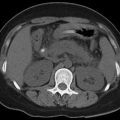
Fig. 23.1
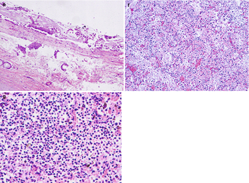
Pathology of measles. (a, b) Slight congestion and edema at the meningeal surface and cerebellar herniation. (c, d) Enlarged volume of both lungs, with edema, multiple ulcers, and blood stains on their surface. (e) Necrosis and shedding of tracheal mucosa. (f) Complete consolidation of lung tissue, with exudation of large quantities of inflammatory cells, destructed tissue, and accompanying hemorrhage. (g) Destructed structure of alveolus, with exudation of a large quantity of cells and proliferation of histiocytes (Note: The figure was provided by Li HJ from Department of Radiology, You’an Hospital, Capital Medical University, Beijing, China)
23.4 Clinical Symptoms and Signs
The incubation period of measles commonly lasts for 6–12 days, with a mean of about 10 days. It can be prolonged to 3–4 weeks in the cases with a history of passive or active immunization.
23.4.1 Typical Measles
The clinical course of typical measles can be divided into three stages.
23.4.1.1 Prodromal Stage
The prodromal stage lasts for 3–5 days from fever to eruption of skin rash. This stage is characterized by upper respiratory inflammation and catarrh symptoms caused by conjunctivitis. The patients experience an acute onset, with fever, cough, rhinorrhea, lacrimation, conjunctiva congestion, photophobia, sore throat, and fatigue. In some cases, the patients might experience headache and gastrointestinal symptoms such as vomiting and diarrhea. During days 2–3 of the illness course, Koplik’s spots occur in above 90 % of the patients with measles, a characteristic sign of measles at its prodromal stage with diagnostic value. In the prodromal stage, some patients may experience rubella-like rashes at the neck, chest, and abdomen, but it is transient lasting for about several hours. These rashes are known as prodromal rash of measles.
23.4.1.2 Eruptive Stage
After days 3–5 of the illness course, fever and respiratory symptoms exacerbate obviously, with eruption of skin rashes. The rashes firstly occur behind the ears and on the hair line and gradually spread to the forehead, face, and neck. From top down, the rashes gradually develop to the chest, abdomen, back, limbs, and finally palms and soles. Within 2–3 days, the rashes can be found across the whole body. Firstly, they are light reddish maculopapules, with the color fading away when pressed and with different sizes. Their diameters range from 2 to 5 mm, with normal skin between rashes. At the peak of eruption, the rashes may fuse in dark color. In some cases, the patients experience hemorrhagic skin rashes with no fading of their color when pressed. Along with erupting reaching its peak, systemic symptoms of viremia occur, with a body temperature of even 40 °C. The patients also experience drowsiness or irritation, even delirium and convulsions, exacerbated cough, red pharynx, dry tongue, red and swollen conjunctiva, and photophobia. By physical examination, the patients are found with enlarged superficial lymph nodes, enlarged liver and spleen, dry or moist rales at the lungs, and even heart failure. The adult patients often experience more severe toxic symptoms than the children patients, but rarely with complications.
23.4.1.3 Convalescence Stage
After eruption reaches its peak, the conditions are commonly improved within 1–2 days, with decreased body temperature and obviously alleviated general symptoms. The skin rashes then fade in a sequence of their eruptions, with light brown pigmentation plaques that are absent after 1–2 weeks tiny branny desquamation.
The whole illness course in patients with no complications lasts for 10–14 days. During the course of measles, respiratory symptoms are the most prominent, such as rhinitis, pharyngitis, bronchitis, and pneumonia. The same lesions at the respiratory mucosa may be found at the intestinal mucosa. In the cases complicated by encephalitis, the brain tissue is subject to congestion, edema, spots of hemorrhage, or demyelination. In addition, during the infection by measles virus, the human body is subject to obviously weakened immune responses, which can temporarily relieve eczema, asthma, and nephrotic syndrome (NS). However, the patients are susceptible to secondary bacterial infection and relapse or exacerbation of tuberculosis.
23.4.2 Atypical Measles
Clinically, atypical measles can occur due to some factors, such as variety in age of infected people, immunity of the body, virulence, and the number of viruses which invade the body.
23.4.2.1 Mild Measles
Mild measles commonly occurs in people with partial immunity, such as babies aged before 6 months, people who have received passive immunity recently, and those who have ever vaccinated measles. Mild measles is characterized by fever at a low degree and in short time, sparse and light rashes, atypical or no oral spots, and mild symptoms of respiratory tract. Generally, no complications occur with a course lasting for about 1 week. Immunity acquired after infection of atypical measles is the same as that of typical measles.
23.4.2.2 Severe Measles
Severe measles commonly occurs in patients who have poor body conditions and weak immunity or is secondary to severe infection with high mortality rate.
Toxic Measles
Toxic measles is characterized by severe toxic symptoms and high fever reaching to above 40 °C during onset, accompanied by tachypnea, cyanosis, fast heartbeat, even delirium, convulsion, and coma.
Shock Measles
In addition to toxic symptoms, circulatory failure or heart failure can occur due to shock measles. It is characterized by paleness, cyanosis, peripheral coldness, weak heart sound, fast heartbeat, and decreased blood pressure. Rashes caused by shock measles are dark and sparse or fade suddenly as soon as rashes erupt.
Hemorrhagic Measles
Rashes are hemorrhagic, thus forming suggillations. They don’t fade when pressed. Hemorrhagic measles might accompanied by hemorrhagic viscera.
Herpetic Measles
Rashes are herpetiform and integrated into bullae. Herpetiform measles is characterized by high fever and severe toxic symptoms.
23.4.2.3 Adult Measles
Adult measles is characterized by serious general symptoms, simultaneous or later occurrence of Koplik’s spots, as well as rashed and their late fading. There are many rashes and fewer complications. But pseudo encephalitis might occur in severe cases.
23.4.2.4 Atypical Measles Syndrome
Abnormal measles mainly occurs in patients who contact other patients with measles again 4–6 years after their vaccination to inactivate measles. Abnormal measles is characterized by sudden high fever, headache, myosalgia, stomachache, and no Koplik’s spots. 2–3 days after infection of abnormal measles, rashes erupt and then gradually extend from distal limbs to the trunk. Rashes are polytypic, often accompanied by acral edema. Upper respiratory catarrh symptoms are not evident but with rales in the lungs and swollen liver as well as spleen. Abnormal measles is severe but limited. High measles hemagglutination inhibition antibodies titer during rehabilitation stage and negative viral isolation are the most important diagnostic evidence. It is generally acknowledged that abnormal measles is noninfectious.
23.5 Measles-Related Complications
23.5.1 Central Nervous System
Encephalitis related to measles includes acute encephalitis, immunosuppressive encephalitis, and subacute sclerotic panencephalitis. It is commonly acknowledged that encephalitis related to measles is caused by direct invasion of measles virus to brain tissue. However, the role of immune mechanism cannot be excluded.
23.5.1.1 Measles Complicated by Acute Encephalitis
Measles complicated by acute encephalitis commonly occurs at days 2–6 after eruption. Its clinical manifestations resemble to those of encephalitis, including fever, headache, vomiting, convulsion, drowsiness, and even coma. In addition, meningeal irritation sign positive, increased total cell count in the cerebrospinal fluid with mainly lymphocytes, and occasional high levels of protein and sugar can be observed. According to literature report, transient EEG abnormalities can be observed in about 50 % of the patients with measles in the advanced stage, while increased lymphocytes in cerebrospinal fluid can be found in about 10 % of the patients. However, encephalitis can be clinically observed in only 0.1–0.5 % of the patients. The severity of symptoms has individual differences. Mild encephalitis can be cured within several days, but in rare cases, the disease progresses drastically and rapidly, leading to occurrence of death.
23.5.1.2 Measles Complicated by Immunosuppressive Encephalitis
Measles complicated by immunosuppressive encephalitis is also known as delayed measles encephalitis, which commonly occurs in patients with measles and compromised immunity. At the early stage, measles is characterized by mild and atypical symptoms. However, in the asymptomatic stage that is 2–5 months after the fading of measles, neurological symptoms occur, such as mental disorder, lethargy, coma, convulsion, and hemiplegia. The disease has an acute or subacute onset with a short illness course, with occurrence of death in several weeks to several months after the onset. Experimental studies have demonstrated that measles virus might mutate in the human body with compromised immunity. They achieve higher affinity to the central nervous system and cause pathological changes in nervous tissues. By autopsy, the brain commonly appears normal, with diffusive pathological changes at the brain and spinal cord. Inclusion bodies can be found at the cytoplasm and nucleus of neurocytes as well as in the astrocytes. A large quantity of neurocytes is subject to degeneration and microglia, and astrocytes are subject to large quantities of proliferation. The hippocampal pyramidal layer is subject to severe hypoxia. But the white matter remains normal. Occasionally, infiltration of monocytes around the meninges and blood vessels can be observed. Concerning the case report, one case was reported to develop immunosuppressive measles encephalitis 5 years after kidney transplantation, whose diagnosis was defined by immunofluorescence and electron microscope. By reviewing related literatures, a total of 24 cases have been reported, with 2 adults and 22 children. Such type of encephalitis occurs during the therapies of cytotoxin, hormone therapy, intrathecal chemotherapy, central nervous system radiotherapy or organ transplantation for treatment of acute lymphoblastic leukemia, other neoplasms, and nephropathies. Among the 24 cases, 18 of them had a medical history or contact history of measles. Generally, it occurs 2–5 months after measles is cured, with an acute or subacute onset. The conditions develop from unconsciousness to coma, with confined or systemic seizures, tetany, hemiplegia, and other symptoms.
23.5.1.3 Subacute Sclerotic Panencephalitis (SSPE)
Subacute sclerotic panencephalitis (SSPE) is a long-term complication of measles, which commonly occurs 2–17 years (averagely 7 years) after the occurrence of primary measles. It can be categorized into chronic or subacute progressive encephalitis, which rarely occurs with an incidence rate of 1–4 per million people. Its pathomechanism is related to mutation of viral genes. After viral mutation, the infected human body cannot produce antibodies against the matrix proteins, resulting in long-term incubation of the virus in the brain cells. The patients gradually experience intelligence disturbance; changes of personality; uncoordinated movements; disorders of language, vision, and hearing; as well as epileptic seizure. Consequently, the patients may die from coma and tonic paralysis.
23.5.2 Pneumonia
Measles complicated by pneumonia commonly occurs in children aged below 5 years or adults in poor health. The etiological factors of measles can be divided into two types: bacterial and viral. The common pathogenic bacteria include Staphylococcus aureus, Streptococcus pneumoniae, Streptococcus, and Haemophilus influenzae, while the pathogenic viruses include respiratory syncytial virus and adenovirus. The changes are pathologically characterized by interstitial inflammation induced by virus and infiltration of bacterial alveolitis, including hemorrhage and edema of pulmonary tissue, infiltration of inflammatory cells, formation of microabscesses, and necrosis of bronchial and alveolar walls. The inflammatory exudates extend along the alveolar pores and damaged alveolar walls, with fusion of consolidated lesions. Clinically, the disease is characterized by sudden exacerbation of the conditions and coughing with thick sputum. The pediatric patients may experience flaring of the nasal wings, lip cyanosis, and increased rales at the lungs.
23.5.3 Laryngitis
Laryngitis commonly occurs in children aged below 2–3 years. In the cases with secondary bacterial infection, the larynx is subject to edema and increased secretion to cause laryngeal obstruction. Laryngitis is clinically characterized by voice hoarseness, barking-like cough, dyspnea, and hypoxia. In severe cases, tracheostomy should be performed as early as possible.
23.5.4 Myocarditis and Cardiac Dysfunction
The patients with measles aged under 2 years are susceptible to myocardial diseases, with manifestations of shortness of breath, irritation, pale complexion, and cyanosis. By auscultation, low and blunt heart sounds can be heard, with rapid heartbeat. ECG demonstrates changes of T wave and ST segment.
23.5.5 Others
Measles is also commonly complicated by stomatitis, otitis media, mastoiditis, and gastroenteritis, which are mostly secondary bacterial infection.
23.6 Diagnostic Examination
23.6.1 Laboratory Test
23.6.1.1 Serological Test
Serum-specific IgM and IgG antibodies can be detected, which has favorable sensitivity and specificity and has value for early diagnosis. Detection of serum-specific IgM antibody is the golden standard for the diagnosis of measles.
23.6.1.2 Etiological Examination
Isolation
Secretions from the eyes, nose, and pharynx as well as blood or urine should be collected from the patients at the early stage for inoculation into primary human embryonic renal cells. After the culture, measles virus can be isolated. Such an examination should not be performed as a routine examination.
Detection of Viral Antigen
Nasopharyngeal secretions, blood cells, and cells in the urinary sediment should be collected from the patients to detect antigen of measles virus by immunofluorescence or ELIZA. These specimens can also be used for smear to detect the multinucleated giant cells for the diagnosis.
Nucleic Acid Test
RNA of measles virus can be amplified from clinical specimens by reverse transcription-polymerase chain reaction (RT-PCR). It is also a highly sensitive and specific way for the diagnosis of measles. The test is especially valuable for immune-compromised patients who cannot produce specific antibody.
23.6.2 Diagnostic Imaging
23.6.2.1 X-Ray
X-ray is commonly used for diagnosis of measles complicated by pneumonia.
23.6.2.2 CT Scanning
CT scanning is commonly used to detect measles complicated by pneumonia and encephalitis.
23.6.2.3 MR Imaging
MR imaging is commonly used for the diagnosis of measles complicated by encephalitis, immunosuppressive encephalitis, and subacute sclerotic panencephalitis.
Ultrasound and nuclear medicine are not used for the diagnosis of measles. However, application of PET in studies of subacute sclerotic panencephalitis has been reported in recent years.
23.7 Imaging Demonstrations
23.7.1 Acute Measles Encephalitis
23.7.1.1 CT Scanning
CT scanning demonstrates no abnormality at the early stage. However, several weeks later, CT scanning demonstrates extensive mild atrophy of the cerebral cortex, which is more prominent at the frontal and parietal lobes. And in rare cases, confined hypothalamus atrophy is demonstrated.
23.7.1.2 MR Imaging
MR imaging demonstrates no abnormality at the early stage. However, several weeks later, MR imaging demonstrates mild atrophy of the cerebral cortex, hydrocephalus, and demyelination. The lesions are more commonly found at the white matters around the cerebral ventricles, the basal ganglia, the hippocampus, as well as subcortical and cerebellar white matters, with spots or flakes of long T1 long T2 signal and high FLAIR signal. The lesions are more favorably demonstrated by T2WI. DWI demonstrates limited perfusion. Enhanced imaging demonstrates enhancement of some lesions (Fig. 23.2).
Case Study 1
A male patient aged 29 years complained of fever and rashes for 8 days as well as blurry vision for 1 day. By physical examination, both pupils were equally round with equal size, and the diameter of pupils was 5 mm, direct and indirect light reflex absent, and blindness. He also had red pharynx and one degree enlargement of the tonsils, Koplik’s spots positive, neck rigidity positive, and bilateral Babinski sign suspected positive. By laboratory test, LY% was detected to be 78.9 %.
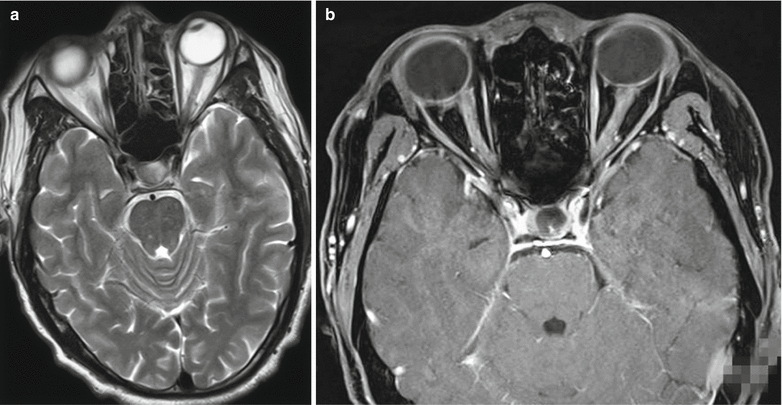
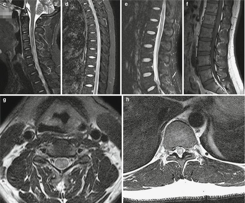
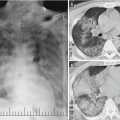
Fig. 23.2
Acute optic nerve encephalomyelitis. (a) Transverse MR imaging demonstrates multiple spots of long T2 signal at the brainstem. (b) Thickened bilateral optic nerves are demonstrated, with uneven enhancement. (c–f) Sagittal imaging demonstrates swelling of spinal cord at the level of C2–T11, with stripes of long T2 signal in the spinal cord. (g, h) Transverse imaging demonstrates long T2 signal in the spinal cord
In patients with spinal cord-related symptoms, MR imaging demonstrates spinal lesions in 80 % of the patients, which are focal or segmental. However, most cases are demonstrated with lesions in long spinal segment (above 3 vertebrae) or whole spinal cord.
The cases of measles complicated by optic neuritis are occasionally reported, with no specificity. Coronal T2WI demonstrates long T2 signal from the optic nerves. Contrast imaging demonstrates enhancement of the lesions, which is more commonly found at unilateral optic nerve.
23.7.2 Immunosuppressive Measles Encephalitis
23.7.2.1 CT Scanning
CT scanning demonstrates multiple patches of low density shadows at the basal ganglia and interface between gray and white matters. Contrast scanning demonstrates no or slight abnormal enhancement. CT scanning demonstrates apparent atrophy of the cortex or even the brain in the advanced stage.
23.7.2.2 MR Imaging
T2WI and FLAIR demonstrate multiple flakes of high signals with poorly defined boundaries. The lesions might involve the thalamus, basal ganglia, bilateral semiovale centrum, interface between gray and white matters, cerebellum, brainstem, and spinal cord. Contrast imaging demonstrates enhancement of cerebral lesions in rare cases and asymmetric lesions at the bilateral brains. The spinal cord can be demonstrated as stripes of long T2 abnormal signal in segmental spinal cord. Contrast imaging demonstrates slight abnormal enhancement.
23.7.3 Subacute Sclerotic Panencephalitis (SSPE)
23.7.3.1 CT Scanning
CT scanning demonstrations are commonly nonspecific, with multiple patches of low-density lesions at the paraventricles, cortex, and white matter. Contrast scanning demonstrates no enhancement. Total brain atrophy might be demonstrated at the advanced stage.
23.7.3.2 MR Imaging
MR imaging is so sensitive to the lesions of SSPE that it is the main neuroimaging examination for its diagnosis.
The lesions are demonstrated to be located at the corpus callosum, basal ganglia, thalamus, and brainstem, especially in the cerebral cortex as well as subcortical and paraventricular white matter. The involvement of white matters might extend to splenium of corpus callosum.
The variety of lesion distribution demonstrated by MR imaging depends on the time of probe. 3–4 months after the occurrence of clinical symptoms and signs, MR imaging demonstrates multiple patches of low T1WI signal and high T2WI signal at the cortex and subcortical white matter. The lesions are commonly located at the parietal and temporal lobes. However, the lesions might involve basal ganglia with symmetric or asymmetric distribution in 20–35 % of the pediatric patients. At the early stage, the cerebral midline structures are subject to shift due to brain tissue edema with space occupying effect and contrast enhancement resembling to neoplasm. With progress of the conditions, the high T2WI signal can extend to paraventricles and corpus callosum. In the advanced stage, high signal lesions at the brainstem can be demonstrated by T2WI, with diffuse cerebral atrophy and occasionally accompanying cerebellar atrophy. SSPE with chronic progress can be demonstrated only as cerebral atrophy. DWI demonstrates slight confined perfusion of the lesions. MRS demonstrates obviously decreased peak of NAA, increased peaks of Cho and mI, observable peak of Lac. Currently, there are no literature reports about application of PWI in studying microcirculatory changes induced by inflammation of brain tissues in the cases of SSPE.
Related posts:

Stay updated, free articles. Join our Telegram channel

Full access? Get Clinical Tree


