Fig. 26.1
Rabies encephalopathy. (a) At day 2 after admission, FLAIR demonstrates high signals in bilateral thalamus, caudate nucleus, and putamen. (b) FLAIR demonstrates high signal of brain gray matter (Reprint with permission from Mani J. Postgrad Med J, Mani, Reddy, Borgohain et al. 2003, 79 (932): 352)
Case Study 4
A boy aged 5 years, with a history of dog bite at the right lateral ankle and no following wound management. He then received 3 successive injections of rabies vaccine. Since 5 days ago, he had high fever, irritation, and gradual inability to walk, with no fears to water, wind, and sound. Based on the findings of pathological changes by autopsy and his clinical manifestations, the diagnosis was made to be paralytic type of rabies.
For case detail and figures, please refer to Yang, N. et al. Journal of Rare Diseases, 2006, 13 (4): 26 (In Chinese)
Case Study 5
A male patient aged 50 years, with manic type of rabies. He had a history of dog bite at the left wrist 7 weeks ago.
For case detail and figures, please refer to
Laothamatas J, et al. Am J Neuroradial, 2003, 24 (6): 1102.
Case Study 6
A patient with rabies (Fig. 26.2).
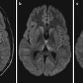
Fig. 26.2
Rabies encephalopathy. (a) T1WI of MR imaging demonstrates multiple spots of low signals in bilateral basal ganglia. (b) DWI demonstrates strips of high signal in the cortex of right frontal lobe (Note: The case and images are provided by Gao, B at Department of Diagnostic Imaging, Yuhuangding Hospital, Yantai, Shandong, China)
Rabies virus is a neurotropic virus, mainly spreading via dog bites in China. It gains its access into the local tissues of human body via wound of dog bite to replicate. After its invasion into the dorsal root ganglion, it replicates in a large quantity to invade the brain and spinal cord, predominantly involving the cerebellum and brainstem, to cause viral encephalomyelitis with a variety of neurological symptoms. Based on the clinical manifestations of rabies, it can be divided into two types, manic type and paralytic type, with the manic type being more common in clinical practice. The clinical manifestations of manic type of rabies include excitement, pharyngeal muscle spasm, paralysis, and hydrophobia, with hydrophobia, ancraophobia, salivation, and mania as its typical symptoms, accounting for over 80 % of clinical rabies cases. The other type of rabies has early manifestations of spinal cross-sectional lesions including progressive flaccid paralysis of limbs and defecation dysfunction, but no symptoms of excitement and hydrophobia, which is known as paralytic type of rabies or dumb type of rabies. In recent years, neuroimaging data of several cases viral encephalitis caused by rabies virus has been reported internationally, which has demonstrations of abnormal MRI signals in the thalamus, hippocampus, caudate nucleus, putamen, and gray matter of dorsal brainstem, with occasional involvements of hypothalamus and pituitary gland. In the cases of a variety of viral encephalitis, including Japanese encephalitis, adenovirus encephalitis, and herpes simplex virus encephalitis, in addition to the predominant pathological changes in the basal ganglia and brainstem nuclei that are the same as rabies virus encephalitis, there are also slight pathological changes of gray matter and white matter as well as diffusive intracranial bleeding. However, intracranial bleeding is rarely found in the cases of rabies virus encephalitis. The characteristic demonstrations by head MR imaging of rabies provide persuasive evidence for the early diagnosis of rabies. Compared to time-consuming etiological examinations, such as virus isolation, detections of antigen and antibody, and animal inoculation, MR imaging can noninvasively demonstrate morphological, functional, and metabolism changes of the brain, which greatly facilitates the early diagnosis of rabies. In addition, it facilitates the differential diagnosis of rabies virus encephalitis from acute disseminated encephalomyelitis and other encephalitis.
Related posts:

Stay updated, free articles. Join our Telegram channel

Full access? Get Clinical Tree


