Medical Techniques and Patient Care

Chapter 5


Medical Techniques and Patient Care


Sonographers work under the direction of a physician-sonologist to obtain diagnostic images of the patients entrusted to their care. Sonography training teaches how to operate ultrasound instruments and to decipher echo information returning from the patient’s body. Sonographers also should know how to provide basic care to patients. The current standard of care recommends using Standard/Universal Precautions in all direct patient contact activities (e.g., wearing gloves whenever in direct patient contact). However, sonographers who find themselves in settings in which strict Standard Precautions are not practiced must wash hands before and after direct patient contact, whenever gloves are not worn or are unavailable.


A sonographer’s obligations


Fulfilling the role of sonographer requires responsibility to yourself, to a department or institution, to patients, and to the profession. From a physical standpoint, sonographers must be adequately rested and relaxed, practice good nutrition, and engage in exercise to promote their physical health. Maintaining good mental health also is important and requires recognizing personal needs, strengths, and limitations. Recognize any anxiety or distress felt about any work-related situations that may interfere with job performance. Leaving personal or family problems at home is just as important as leaving work problems at work. A sonographer who is unable to resolve problems in either area should seek proper counseling. Because caring for patients can be emotionally draining, have a means of rejuvenating through physical exercise and enjoyable hobbies. Developing a good self-image and viewing problems as challenges or opportunities rather than stumbling blocks make it easier to accept criticism as a learning opportunity instead of as a defeat. This kind of attitude brings with it a sense of pride in your work and an eagerness to start each day.


Patient-Focused Care


The most important facet of being a sonographer is seeing the patient as the primary focus of your efforts. Despite personal or philosophic concerns, you must be considerate of your patient’s age, cultural traditions, personal values, and lifestyle. Good patient care goes beyond procedural skills. It includes communicating with patients and allowing them to express their individual problems, fears, and frustrations. Good patient care also requires you to cooperate with other departments and facilities and health care professionals to deliver the best and most complete patient care through a team effort.


Patient-focused care (PFC) represents a national movement to recapture the respect and goodwill of the American public. It is the beginning of a larger objective to ensure that every patient receives the best possible medical care. The patient-focused approach encourages sonographers to relate to patients as people with needs, who are to be respected and cared for in a mature and dignified manner.


Patient rights


In addition to having the necessary knowledge and skills to perform competent sonographic examinations, sonographers are responsible for the type of care given to patients. In 1973 the American Hospital Association (AHA) first adopted a Patient’s Bill of Rights with the expectation that hospitals and health care institutions would support these rights in the interest of delivering effective patient care. The AHA encouraged institutions to translate or simplify the bill of rights to meet the needs of their specific patient populations and to make patient rights and responsibilities understandable to patients and their families. The federal government also was concerned about this issue. In 1998 the U.S. Advisory Commission on Consumer Protection and Quality in the Health Care Industry adopted a Consumers’ Bill of Rights and Responsibilities (also known as the Patient’s Bill of Rights). By 2004 the AHA had replaced the Patient’s Bill of Rights with a plain language brochure, The Patient Care Partnership: Understanding Expectations, Rights and Responsibilities, which informed patients about what to expect during their hospital stay with regard to their rights and responsibilities. The most recent update took place in 2008 and includes translations in English, Arabic, Simplified and Traditional Chinese, Russian, Spanish, Tagalog, and Vietnamese. It is available at http://www.aha.org/aha/issues/Communicating-With-Patients/pt-care-partnership.html.


Health care providers know that all patients deserve to be treated with respect, dignity, and kindness. As health care professionals, sonographers also must remember that patients’ needs come first. Learn to control any anger or frustration that may develop when working with patients. Patients have the right to information concerning their health care and to decision making regarding their diagnosis and treatment. In some instances, patients may refuse care or tests. In such cases, sonographers may try tactfully to reason with them by explaining the nature of the examination as it relates to their problem or illness. Patients who continue to refuse an examination should be encouraged to contact the physician for a fuller explanation. Sonographers should provide reassurance that they gladly will reschedule the examination at a mutually convenient time if the patient decides to undergo the procedure. Sonographers are expected to discuss matters pertinent to patients only with authorized hospital personnel.


The Affordable Care Act


On March 23, 2010, the Affordable Care Act was signed into law as a way of putting consumers back in charge of their health coverage and care. The goal of the act was to crack down on some of the most egregious practices of the insurance industry while providing stability and flexibility to families and businesses. The Departments of Health and Human Services, Labor, and Treasury issued regulations to implement a new Patient’s Bill of Rights under the Affordable Care Act to help patients with pre-existing conditions gain coverage and keep it, to protect patients’ choice of doctors, and to end lifetime limits on the care consumers may receive. Other benefits of the Act are to strengthen review of insurance premiums; to get the most from premium dollars; to keep young adults covered; and to provide affordable coverage to consumers without insurance, because of pre-existing conditions. The new Patient’s Bill of Rights regulations now include the following stipulations:


More detailed information can be obtained by accessing http://www.healthcare.gov/.


Health Insurance Portability and Accountability Act (HIPAA)


The HIPAA Privacy Act was first signed into law in 1996 and created national standards to protect individuals’ medical records and other personal health information. In April 2001 the Department of Health and Human Services enacted the Health Insurance Portability and Accountability Act (HIPAA). The HIPAA Privacy Rule became effective April 14, 2003, marking the first comprehensive federal protection for the privacy of health information. Pursuant to this act, all segments of the health care industry (including sonography) are charged with promoting enhanced patient privacy in the health care system. The HIPAA Security Rule, which required full compliance by April 21, 2005, is a federal law, and anyone not in compliance can face up to $250,000 in fines and jail time of up to 10 years.


The Privacy Rule was created to achieve the following:


• Give patients more control over their health information


• Set boundaries on the use and release of their health records


• Establish appropriate safeguards that health care providers must achieve to protect the privacy of health information


• Hold violators accountable, with civil and criminal penalties that can be imposed for violation of patients’ privacy rights


• Strike a balance when public responsibility requires disclosure of some forms of data (e.g., to protect public health)


• Enable patients to find out how their information may be used and what disclosure of their information was made


• Limit the release of information to the minimum reasonably needed for the purpose of the disclosure


• Give patients the right to examine and obtain a copy of their own health records and to request corrections


Sonographers must understand the privacy procedures of their practice or hospital to be able to provide patients with information and answers to questions about how their information can be used. Sonographers also are tasked with securing patient records containing individually identifiable health information so that they are not readily available to those who do not need them. Any communications necessary for quick, effective, high-quality health care are considered allowable communications. These include communicating verbally with pertinent physicians and family members and using patient names to locate them in waiting areas. In compliance with HIPAA, sonographers must be careful to do the following:


Patients may request that students, other observers, medical personnel, and families leave the room during the sonography examination. If any patients elect to have friends or family members present during their examination, inform them of the hospital rules and any policies that govern such requests. Some sonographers say they feel uncomfortable having “outsiders” looking on while they are scanning, even though no regulations may prohibit their presence. If this situation arises, give a truthful explanation to the patient if inadequate space exists for viewing or if the presence of observers hinders your concentration and abilities. Explain that you need time alone with the patient to prepare for the examination and to take a series of scans for evaluation purposes. This may ease the situation. In return for such cooperation, you could offer to call the observers into the examination room for their own “viewing.” By following these suggestions, you can control the discovery of unexpected pathologic findings without causing anxiety to patients or their support group.


Another patient right involves the expectation of pleasant physical and emotional surroundings that ensure the person’s comfort, safety, and respect as an individual.


Patient environment


One of the most important considerations in the design of an ultrasound facility should be the patient’s and the sonographer’s physical surroundings. For safety and comfort the following features should be considered:


Usually, the sonographer’s duty is to keep the examining room and equipment neat and clean. Properly clean any nondisposable medical equipment used by the patient immediately after an examination and return it to its proper place. Contact housekeeping personnel for immediate services in case of accidental spills.


Emotional Surroundings


An ultrasound facility should offer a climate in which the patient is treated as an individual. Staff members should introduce themselves to patients and explain what they will be doing for them. Patients should be oriented to any procedures such as filling out admission or consent forms. Patient privacy should be respected at all times, particularly during dressing and undressing, during performance of ultrasound scans, and during use of bathroom facilities. If patients require assistance at any of these times, the sonographer should provide it in a mature and completely professional manner.


Comforting patients is at least 80% nonverbal and involves giving the patient undivided attention, using facial expressions, eye contact, gestures, and voice intonations that convey empathy.


A sonographer should allow patients to freely express their thoughts, opinions, or beliefs. Be a good listener and do not impose beliefs on the patient. In the event that the examination performed reveals serious illness or the threat of imminent death, patients may want to engage in spiritual practices or rituals. Show respect for their wishes and provide assistance or privacy as indicated.


Patient care


The Spectrum of Patient Reactions to Illness


Sonographers are in a position to see the changes that disease and disability cause in people. Any significant change is a series of gains and losses as disease and disability interrupt the natural balance of the body. These changes produce stress, and such stress causes emotional reactions in patients.


Learning of illness brings about varied but powerful reactions in patients. Emotions may range from shock to relief and everything in between. Even if the patients’ symptoms have been evident for a long time, hearing the diagnosis may be upsetting. Also daunting to patients are illnesses that include periods of good health mixed with periods of sickness. Constantly having to readjust to these changes can take a toll. The following are among the important emotional reactions a sonographer may encounter.


Suspicion


Feelings of mistrust may overcome some patients, making them fearful and feeling that everyone and everything is against them. Often, the one-on-one environment of the sonography laboratory encourages patients to talk about their feelings. Patients who finally reach the stage of acceptance understand that they have a right to be emotional but that that behavior ultimately robs them of taking control through knowledge, problem solving, and cooperating with their health care team. Emotional relapses may occur, but they will learn to recognize and move past them.


Although sonographers are not responsible for evaluating and treating patients’ emotional reactions, they should share observations and concerns with the referring physician or charge nurse. By all means, try to be a good listener. If a patient responds negatively to being scanned and becomes upset, do not also get upset. Instead, try to be patient, understanding, and secure enough to let patients know you care.


Helping Patients Understand How to Cope with Their Illness


When dealing with illness—especially chronic illness—patients find strength they never thought they had. Acceptance of their condition is essential, along with finding ways to feel more in control. Sonographers can play a part in helping their patients take control by discussing how knowledge can help them understand what is being done to them, make better decisions, and be proactive. Working with the health care team by being cooperative ultimately will help them find answers. Learning how and when to relinquish some control can only speed the quest for answers and recovery. Sonographers who spend extended time with such patients can gauge the level of their concern. They have the opportunity to discuss how patients may gain hope and understanding through support networks, through sharing concerns and fears, and through learning to ask for and accept help.


Vital signs


The term vital signs refers to temperature, pulse, respiration, and blood pressure as indicators of the functioning of the body. During the course of a sonographic examination, sonographers may be required to assess the pulse, respiration, and blood pressure as part of the scanning protocol. Careful, accurate measurement of each of these parameters is essential.


As a part of the assessment of vital signs, the sonographer should observe the patient’s total condition: color, skin temperature, and patient feelings and reactions. Sonographers who have never worked with patients before will benefit from the following review of the vital signs, which will establish guidelines for obtaining and understanding their significance.


Pulse


The pulse is the beat of the heart that can be felt as a vibration within the walls of the arteries. With each heartbeat, blood forced into the arteries causes them to swell or expand, producing arterial pulses that can be felt with the fingers. The most convenient site for taking the pulse is the radial artery, located on the thumb side of the wrist. However, other arteries close to the skin also provide pulse sites (e.g., temporal, carotid, mandibular, femoral, and popliteal).


Pulse rate refers to the number of beats per minute (bpm), whereas rhythm refers to the time interval between beats. The sonographer should evaluate whether the pulse has a smooth and regular rhythm as opposed to irregular rhythms with skipped beats. The strength of a pulse refers to its force and usually is described as either bounding or weak and thready. Substances such as coffee, tea, tobacco, or certain drugs can cause rhythm irregularities. Shock and hemorrhage can cause a weak, thready pulse, whereas fever can produce a bounding pulse. The normal adult pulse rate is 60 to 65 beats per minute (bpm). Newborn infants have a pulse rate of 120 to 140 bpm, and women, children, and elderly patients usually have a slightly faster than normal pulse rate (Table 5-1). Athletes in good condition generally have a slower pulse: below 60 bpm.


Any variation of the normal rhythm of the heart is termed arrhythmia and includes premature beats or palpitations. Such irregularities may simply be a normal physical response or a sign of disease. Abnormally rapid pulse rates (> 100 bpm) are termed tachycardia. Abnormally slow pulse rates (< 60 bpm) are termed bradycardia. Exercise, strong emotions, fever, pain, and shock can elevate the pulse. In contrast, resting, depression, and certain drugs (such as digitalis) can lower the pulse.


The pulse is felt by gently compressing the artery over a bony prominence in the area. Never feel the pulse with the thumb, because the thumb has a pulse of its own that interferes with obtaining an accurate reading of the patient’s pulse. An arterial pulse should be obtained as follows (Figure 5-1):
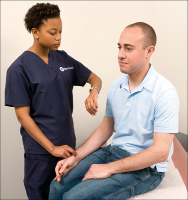


Respiration


The oxygen and carbon dioxide exchange that occurs in the lungs is referred to as respiration. The respiratory process begins with the delivery of oxygen to body cells via blood that has passed through the lungs. The cells give off accumulated carbon dioxide to the blood, which returns it to the lungs. There the potentially dangerous carbon dioxide wastes are exhaled out of the body in the act of breathing.


Breathing can be defined as the expansion (inspiration) and contraction (expiration) of the lungs. Normal breathing is quiet, effortless, and regular in rhythm; it occurs at a rate of 16 to 20 breaths per minute in the normal adult.


Note the rate, rhythm, and the depth and character of the respiration. The rate refers to the number of respirations per minute. Rhythm refers to the regular rate of breathing and a symmetric movement of the chest. Depth refers to the amount of air taken in with each respiration (normal, shallow, or deep). Character refers to the quality of respiration (e.g., quiet, labored, wheezing, and coughing).


Any injuries to the lungs, chest muscles, or diaphragm affect breathing. Note any positions the patient may need to assume to breathe easily (e.g., sitting up or standing as opposed to lying down). Also note any difficulty in breathing (dyspnea) or changes in the patient’s color (cyanosis or pallor).


Count respiration without the patient being aware of this task. Watch the patient’s breathing and count a breathing sequence (in and out) as one respiration. Start the count at zero and continue counting respirations for at least 30 seconds. Multiply the number by 2 to produce the respirations per minute. If irregularities are observed (rate, rhythm, patient appearance, or behavior), count the respirations for 1 full minute and report the findings.


Blood Pressure


Blood pressure is the pressure that circulating blood exerts against arterial walls. It is produced by the pumping action of the heart. The pressure of blood within the arteries is highest whenever the heart contracts (called systolic pressure). Between beats, when the heart rests, arterial pressure is at its lowest (called diastolic pressure).


Heart activity can be heard as “thumping” sounds in the large arteries of the limbs. These sounds can be translated into numbers representing millimeters of mercury (mm Hg) on a manometer. When taking a blood pressure reading, the sonographer listens with a stethoscope and watches the numbers on a manometer.


The optimal adult blood pressure is less than 120/80 (120 indicating systolic pressure; 80 indicating diastolic pressure). High blood pressure, or hypertension, is known as “the silent killer,” because most individuals have no symptoms. It causes wear and tear of the delicate inner lining of the blood vessels. Heredity and aging are the greatest risk factors. The risk begins to increase from pressures of 115/70 mm Hg and doubles for each 10 mm Hg increase in the systolic reading and 5 mm Hg increase in the diastolic reading. Although variations exist even in normal patients, the average values for arterial pressure are as follows:


Even if only one reading (either the systolic or the diastolic reading) is in the high range, it signifies high blood pressure. Strong emotions, pain, exercise, and some disease conditions are factors that can increase blood pressure. Resting, depression, hemorrhage, and shock are factors that can lower blood pressure. Blood pressure readings also can vary from minute to minute and day to day because of changes in a patient’s physical, mental, or emotional activity.


The volume of blood in the body and any resistance to the flow of blood through blood vessels also affect blood pressure. For example, hemorrhaging decreases blood volume, causing the blood pressure to fall. In contrast, fatty deposits develop within the blood vessels of patients with arteriosclerosis, causing resistance to blood flow and producing higher blood pressure readings.


The site for taking blood pressures is usually over a large artery in the arm or leg. The brachial artery of the upper arm is selected often, but arteries of the lower arm, thigh, and calf also may be used, depending on the accessibility of the limb and the patient’s condition.


When the arm is used, the patient should either sit or lie down on his or her back, with the arm and blood pressure cuff at the level of the heart.


A wide variety of blood pressure monitors are available: mercury/aneroid, digital, and multipurpose vital sign monitors. The most familiar of these is the sphygmomanometer (either mercury or aneroid), used with a cuff and a stethoscope (Figure 5-2). Mercury manometers, which provide the most accurate measurements, show increments of 10 points, from 0 to 300. Aneroid manometers show units of 20, ranging from 20 to 300. Raising the silver column of mercury or the aneroid needle to 200 begins the procedure. A properly calibrated manometer will show the needle and mercury column at the zero mark on the mercury manometer (20 on the aneroid manometer) when not in use.
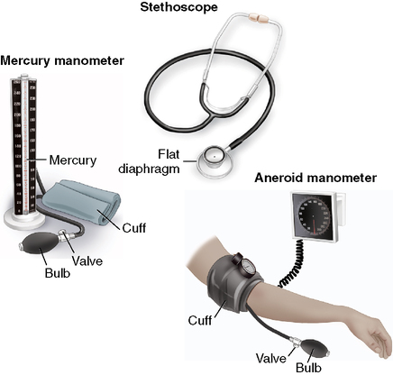


The cuff contains an inflatable rubber bladder; center this over the brachial artery, 1 inch above the elbow (Figure 5-3). Wrap the cuff snugly but not tightly. If the cuff is too loose, systolic and diastolic readings will be heard higher than their actual values.
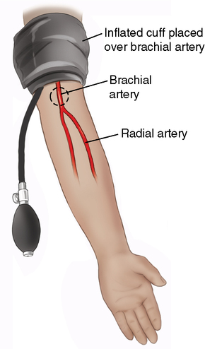


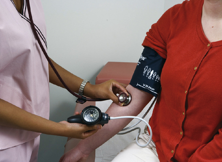
A stethoscope with a flat diaphragm is best for taking blood pressure. After using your fingers to locate the brachial artery in the shallow depression where the elbow bends, place the diaphragm of the stethoscope over the artery without touching the cuff or the patient’s clothing (Figure 5-4). Use gentle application, because too much pressure can cause abnormally low diastolic sounds.


Measuring blood pressure


Sonographers measuring blood pressure should do the following:


• Explain the procedure to the patient in a private and noise-free environment.


• Ensure that the patient rests for 5 minutes before the blood pressure is taken. If the patient has been actively exercising, take the reading after 15 to 30 minutes of rest.


• Select and use the same site consistently, because variation occurs in blood pressures taken in different locations. Take blood pressures with the patient in the same position each time. A lying, sitting, or standing blood pressure can vary within the same individual.


• Remove, as necessary, any of the patient’s clothing, to expose the site. The patient’s arm should be positioned on a supporting structure (table or bed) at heart level while the blood pressure is being taken.


• Check that no leaks exist in the tubes, cuff, or valve, and remember that using too small a cuff can result in pressure readings higher than normal.


• Sit or stand when taking the blood pressure to read the manometer at eye level. The manometer should read 0 before beginning.


• Deflate any air out of the cuff, and apply the cuff just far enough above the patient’s elbow to leave the space over the brachial artery free. Wrap the cuff firmly around the patient’s arm and fasten it securely on the last turn.


• Locate the pulse in the artery and place the stethoscope directly over the point of strongest pulsation.


• Hold the rubber bulb in the palm of one hand; close the valve on the rubber bulb with a thumb and finger and rapidly inflate the cuff by pumping the bulb.


• Inflate the cuff to about 20 to 30 mm Hg above the expected systolic reading. As the cuff pressure increases, it will shut off the flow of blood within the artery. Inflation of the cuff should take 7 seconds or less.


• Carefully loosen the valve and deflate the cuff slowly and steadily. Then listen carefully for sounds of the first heartbeat (systolic pressure) and watch the mercury column or aneroid needle gauge for the points at which the sounds are first heard.


• Deflate about 2 to 3 mm Hg per heartbeat until all sounds stop or a distinct change occurs in the sound (diastolic pressure).


• When all sounds stop, deflate the cuff rapidly and completely to 0.


• Do not reinflate the cuff during the reading. If you must repeat a reading, let all the air out of the cuff and allow 15 seconds to lapse before inflating the cuff again.


If the cuff is deflated too slowly, false elevated readings will result. If the cuff is deflated too quickly (5 to 10 mm Hg/heartbeat), false low readings will be obtained.


Interpreting arterial sounds


Arterial sounds are interpreted as follows:


Distinct changes in the sounds may be heard between the systolic and diastolic sounds. Sounds may become muffled before stopping or may remain muffled down to 0. In this case, record the diastolic at the point when you hear the change from clear to muffled and when the sound ends completely (e.g., 120/76/62). Write the reading down promptly.


Digital manometers were developed for professional use in hospitals, clinics, and doctors’ offices. They provide automatic cuff inflation and take up to three pressure readings before averaging the total for the most accurate measurement. Wrist cuff monitors also are available. Their small size makes them ideal for portable work and for home monitoring practices.


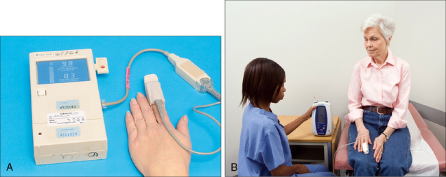
Continuous vital signs monitors also are widely used in hospital settings. They are designed to spot check or continuously monitor several critical parameters such as heart (pulse) rate, pulse oximetry, blood pressure, and temperature. Pulse oximeters are used in various situations, especially in surgery and the intensive care unit (ICU). Pulse oximetry measures the oxygen concentration in arterial blood. The normal range is 95% to 100%. Measurements are used to prevent hypoxia and to evaluate the effectiveness of treatment. A sensor is attached to the patient’s finger, toe, earlobe, nose, or forehead. The sensor light beams pass through the tissues, and a detector on the other side measures the amount passing through the tissues. The oximeter then measures the oxygen concentration from this information (Figure 5-5). That value, and the patient’s pulse rate, are displayed on the monitor. Oximeter alarms will sound if the oxygen concentration is low or if the pulse rate is too fast or too slow. Easily attached to IV poles or on rolling stands, these monitors may accompany inpatients who must be transported to the ultrasound laboratory.


Intravenous Equipment


IV tubing connected to a plastic bag or bottle is used to infuse fluids into the patient’s body. A needle or plastic catheter attached to the container is inserted into a vein. The flow of fluid is measured by a drip meter, and a clamp on the tubing is used to regulate the flow of the prescribed fluid (Figure 5-6).
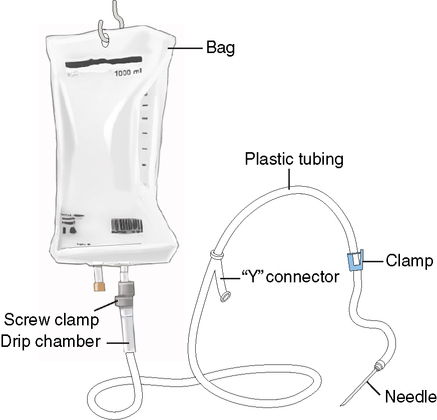


Some institutions may use computerized infusion pumps to regulate drip rate. This device delivers a measured amount of fluid over a period of time. An alarm sounds if a problem occurs, such as drops falling at an improper rate, the bag emptying, infiltration within the patient’s tissues, or patient’s arm position obstructing flow.


Unless specifically authorized, you should not change or regulate the amount of flow of a solution, although you may have to move some patients to and from the scanning table or bed or remove their clothing. The following guidelines should help in working with such patients:


1. If the needle has been inserted in the patient’s hand or arm, help the patient keep the involved arm straight.


2. Never lower the bottle or bag below the level of the needle insertion when transferring or positioning the patient.


3. Watch for and immediately report any of the following:


• Nausea, vomiting, rapid breathing, or an increase in pulse rate occurs, which are signs of circulatory overload that must be dealt with immediately.


• No solution is passing from the bottle into the tubing, even though solution is still in the bottle.


• The plastic drip chamber is completely filled with solution.


• Blood appears in the tubing at the needle end.


• All of the solution has run out of the bottle or bag or it is almost empty.


• The needle has deliberately or accidentally been removed.


• The patient complains of pain or tenderness at the needle insertion site.


• A raised or inflamed area occurs on the patient’s skin or near the needle insertion, which may mean that the solution has infiltrated and is running into the adjacent tissues (the sonographer may be asked to close the clamp to shut off the flow of solution).


• The tubing becomes disconnected, and the patient is bleeding freely from the connection site.


Nasogastric Tubes


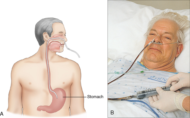
Hospital patients commonly have a tube inserted into one nostril. Depending on the length of the tube, it may terminate in the patient’s stomach or intestine. Such tubes can be used for feeding, to obtain specimens, to treat intestinal obstructions, to prevent or treat distention after surgery, or to drain fluids from the patient’s stomach by suction (Figure 5-7).


When a nasogastric (NG) tube is being used to drain substances out of the stomach or to collect a specimen, the patient is given nothing by mouth (NPO), because food would only be drawn back out through the tube. In caring for patients with NG tubes, never pull on the tube when moving these patients or changing their positions. If the patient begins to gag or vomit while the tube is in place, report it immediately.


Fluids can be removed from the patient’s body through the tubes by gravity or suction. The outer end of the tube may have a clamp attached, or a plastic connection may link it to longer tubing attached to an electrical suction machine. Suction (by use of either a syringe or a machine) often is used to remove thick secretions that cannot be drawn out easily by gravity.


Here are some rules for working with patients connected to mechanical suction machines:


Oxygen Therapy


Oxygen may be administered to any patient experiencing respiratory difficulty (oxygen deficiency). The goal of such therapy is to lessen a low-oxygen concentration in the blood and to decrease the workload of the respiratory system.


The use of oxygen is associated with some hazards. Oxygen should be considered a drug and its dosage or concentration should be evaluated and ordered by a physician. Although oxygen by itself cannot burn, contact with any combustible material (even a spark) causes it to ignite and burn or, in high concentrations, to explode.


Delivery systems for oxygen therapy include in-room piping systems and oxygen tanks, or cylinders. If a laboratory is equipped with an in-room piping system, oxygen and suction usually are provided through wall outlets. Outlet connectors, which vary in shape, color, and connection methods used, are keyed to a specific gas or function (Figure 5-8).
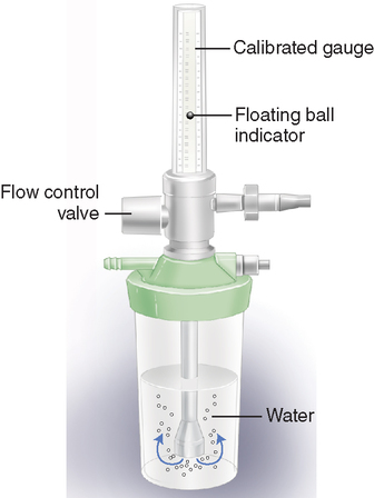


Oxygen also may be contained in large tanks or small cylinders. Large cylinders usually are used for patients requiring high flow rates or oxygen use over extended periods. These can be identified by size and the presence of a metal cap screwed onto the top of the cylinder to protect the valve from damage. Small cylinders can be identified by their size and a rectangular valve (without a handle), which has three holes on one side. Small cylinders are used during patient transportation or for short duration needs. Some ambulatory patients need continuous oxygen and may use over-the-shoulder slings or rolling stands to hold the tank when ambulating.


Because the gas contained within a cylinder is under extremely high pressure, sonographers should never do the following when gas cylinders are in use:


The delivery of oxygen to the patient may involve the use of either high- or low-flow devices. Among the low-flow devices are the nasal cannula or nasal prongs, nasal catheter, or simple oxygen masks. The nasal cannula is used when a patient needs extra oxygen rather than a total supply of oxygen (Figure 5-9, A). The prongs are inserted into the patient’s nostrils and held in place by an elastic band around the patient’s head. They are connected to the oxygen source by a length of plastic tubing.
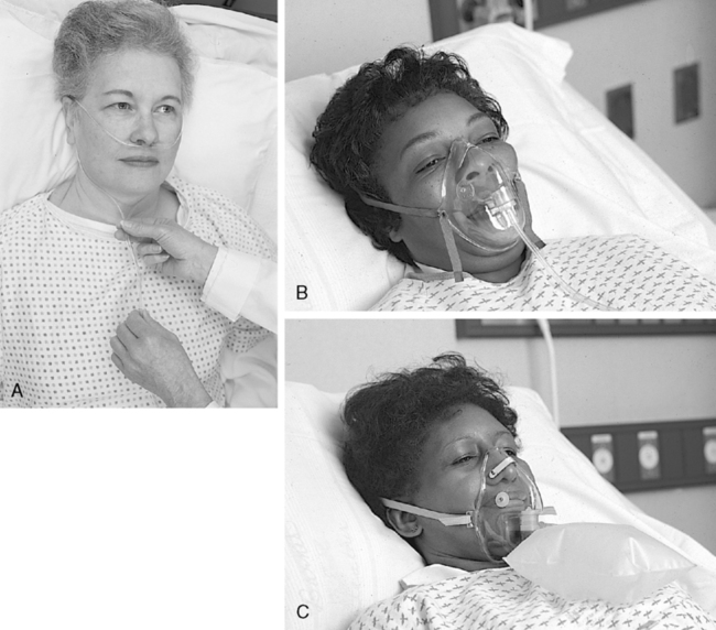


A nasal catheter is a piece of tubing that is longer than a cannula. It is inserted through the nostril into the back of the patient’s mouth. This method provides more effective oxygen delivery and is used when the patient must have additional oxygen at all times. The nasal catheter is fastened to the patient’s forehead or cheek by a piece of adhesive tape to hold it steady.


Several different types of oxygen masks may be used. Among the most common types of masks are the following:


All oxygen masks, except the Venturi mask, require humidification, which is generally achieved by means of a humidifier (usually disposable) filled with water. The humidifier is connected to a threaded outlet at the bottom of the flowmeter or regulator. A small universal connector extends from the front or top of the humidifier for connection to the oxygen device. The following precautions should be observed in working with patients receiving oxygen therapy:


Catheters


The most common kind of catheter used for removing fluids from the body is the urinary catheter. The catheter (made of plastic tubing) is inserted through the patient’s urethra and into the bladder (Figure 5-10). Catheters may be used to obtain sterile specimens when patients are unable to urinate naturally or to determine how much residual urine remains in a patient’s bladder after urination.
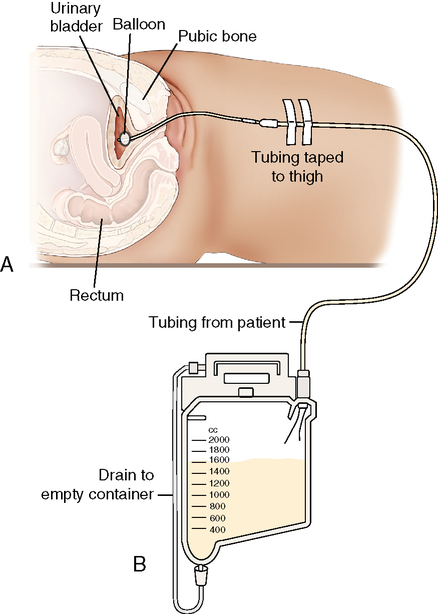


A retention, or indwelling, catheter is a system used to provide temporary or permanent drainage of urine. A Foley catheter commonly is used. Foley catheters are specially designed as two tubes, one inside the other. The inner of the two tubes is connected at one end to a small balloon. After the catheter has been inserted, the balloon is filled with water or air so that the catheter will not pass out through the patient’s urethra. Urine drains from the bladder through the outer tube and collects in a container attached to the patient’s bed or table. Clamping or unclamping the tubing can control the flow of urine. Importantly, the catheter bag must always be positioned at a level lower than the patient’s urinary bladder. Catheterization is a sterile technique, and the catheter drainage system should consist of a closed sterile system. As with other patient tubing, the catheter should be taped to the patient (inner thigh in this case) at all times and should be checked periodically for kinking and to ensure that the patient is not lying on the tubing and obstructing the flow of urine out of the body.


Another use of the urinary catheter involves patients scheduled for obstetric or gynecologic sonography. Some of these patients are unable to fill the bladder naturally or maintain a full bladder for the duration of the examination. As a result, they must be catheterized to instill fluid into the bladder, with the tubing clamped off until the end of the sonography study. Bladder filling is not required for transvaginal examinations.


Because of the short length of the female urethra, the risk of urinary tract infection is increased with catheterization. For this reason, it is unwise to use catheterization as a routine method of filling the bladder of patients scheduled for pelvic sonography, unless a clinical indication exists to do so. Only trained and authorized personnel should perform any catheterization procedure.


Colostomies and Ileostomies


Surgical treatment of patients with disorders of the intestinal tract may result in the construction of a colostomy (an opening into the colon) or an ileostomy (an opening into the ileum). In such patients, a loop of intestine is brought out of the body through a surgical incision on the abdominal wall; the opening allows drainage of feces. The colostomy or ileostomy opening (stoma) is covered by plastic disposable bags or pouches held in place by double-faced adhesive tape or special glue. These appliances may require frequent changing because of the constant flow of liquid feces that is especially prevalent during the immediate postoperative period.


When performing abdominal sonography on such patients, have a small supply of appliances on hand. It may be necessary to remove and later replace the devices to gain unobstructed access to the surface of the abdominal wall. Follow these procedures:


Stay updated, free articles. Join our Telegram channel

Aug 20, 2016 | Posted by in ULTRASONOGRAPHY | Comments Off on Medical Techniques and Patient Care

Full access? Get Clinical Tree

Get Clinical Tree app for offline access