Students who successfully complete this chapter will be able to do the following: • Describe the evolutionary history of diagnostic ultrasound. • Identify at least four of the leading pioneers of diagnostic medical ultrasound. • Compare and contrast the imaging modalities discussed in this chapter. • Discuss the ways in which ultrasound is used as a therapy and for diagnosis. • Describe how hand-carried ultrasound is affecting clinical medicine. • Discuss the differences between 3D and 4D ultrasound. • Explain the special importance of musculoskeletal ultrasound to sonographers. • Discuss why ultrasonic harmonic imaging is superior to conventional ultrasound. • Explain how high-intensity, focused ultrasound differs from conventional ultrasound. The goal of this chapter is to acquaint the reader with some of the significant milestones and pioneers in the history of acoustics and medical ultrasound (Table 1-1). The taproots of diagnostic ultrasound lie in one of the oldest sciences—the science of sound, or acoustics—which dates back to the ancient Greeks. Earlier cultures, such as the Egyptians, Persians, and the Chinese, developed musical instruments and became interested in how sound was created. However, a systematic study of sound really began with the Greek mathematician Pythagoras, thought to have been born around 550 BC. Pythagoras observed the relationship between sound pitch and frequency and is thought also to have invented the sonometer, an instrument used to study musical sounds. The Greek scholar Archytas, a Pythagorean and contemporary of Plato, correctly showed that pitch is related to the movement of vibrating air. However, he incorrectly asserted that the speed at which the vibrations travel to the ear is a factor in determining pitch. It was not until 350 BC that Aristotle developed the theory of sound propagation, the idea that sound is carried to the ears by the movement of air. Boethius compared sound to water (Box 1-1). Table 1-1 Historical Milestones in the Study of Sound: 500 BC to 1883 AD The development of metal flaw detectors and naval sonar also made possible the work of three independent American investigators: George Ludwig, John Wild, and Douglass Howry. Between 1947 and 1949, Ludwig, a Naval medical research surgeon, joined colleagues at the Massachusetts Institute of Technology. Using A-mode studies exclusively, Ludwig was able to detect successfully gallstones embedded in animal tissues (Figure 1-1). Wild, an English surgeon who immigrated to America, was the first to consider using ultrasound to detect tissue thickness. In his work at the University of Minnesota, he realized that ultrasonically, cancerous tissues differed greatly from normal tissues. Along with engineer John Reid, he constructed an early prototype breast scanner that employed the use of an externally placed water path. Their B-mode (brightness modulation) techniques used 2D presentations of echo-producing interfaces (Figure 1-2). In 1955 Wild and Reid presented the first paper on ultrasound used for medical purposes.1 Wild was also a pioneer in the development of early internal scanners and devised a rectal transducer to obtain images of the large bowel. In Denver, radiologist Douglass Howry worked independently of the other groups. Howry had pursued an interest in diagnostic ultrasound since 1948. In 1951, using war surplus electronic components, he constructed a “water-path” scanner. A laundry tub and later a cattle tank were the first prototypes for holding the water in which the subject or the body part to be imaged was submersed in water (Figure 1-3). Unfortunately, the one-dimensional images that resulted were disappointingly incomplete. Howry joined his friend and mentor, Joseph Holmes, who was at the University of Colorado in Denver. Their collaboration resulted in the development of a compound scanner. Howry and Holmes discovered that by simultaneously moving the transducer in two different motion patterns, a more complete anatomic picture could be formed. The cattle tank eventually was replaced by an upturned B-29 gun turret, in which the subject was immersed and weighted down to avoid introducing motion artifacts as the mechanically circling transducer cut a path through the water (Figure 1-4). The impracticality of using this method on sick patients spurred the two scientists to simplify the procedure and to develop a water-bath or “pan” scanner in 1957 that permitted the patient to sit next to a small pan of water through which the transducer moved. Their experience eventually led Howry and Holmes to the development of a compound contact scanner, which provided direct scanning of the body using a light film of oil or lubricating gel to replace the cumbersome water path (Figure 1-5). The early bi-stable images registered on a phosphorus-coated oscilloscope screen as dots of light. All echoes greater than predetermined specific amplitudes were displayed as constant-intensity dots of light. After Howry left Denver to work at the Massachusetts Institute of Technology, Holmes worked closely with William Wright, an electronic engineer from Loveland, Colorado. The fruits of their labor were realized in 1962 with the introduction of the first commercially available portable ultrasonic system, a compound contact scanner known as the Physionics Engineering Porta-Arm (Figure 1-6). This system gained worldwide acceptance and use. The work of the Denver group is considered one of the most seminal pioneering contributions in B-mode imaging and contact scanning. The Porta-Arm scanner was the direct precursor to the imaging systems in use today. In the mid-1950s, obstetrician Ian Donald, aware of Howry’s work and that of Japanese researchers, began his study of diagnostic ultrasonography in Scotland. His interest stemmed from his experiences with the Royal Air Force during World War II, during which time he witnessed the ultrasonic testing of aircraft to detect metal stress and fatigue. Donald vowed to pursue his notion that ultrasound may be used in a similar fashion on patients. By means of comparative examinations of an excised tumor and a beefsteak (from the local butcher shop), Donald proved that tumors possessed echo patterns different from those of normal tissue. This work was conducted in an atomic boiler plant outside Glasgow with use of a borrowed flaw detector. Later, again using borrowed equipment and A-mode technique, he began detecting ovarian cysts, ascites, and polyhydramnios in his patients. Donald is credited with perfecting the first A-mode measurement of the fetal biparietal diameter, making it possible to ultrasonically estimate fetal age, weight, and growth rate (Figure 1-7). A few months earlier, on October 29, 1953, equipped with the Siemens Ultrasound Reflectoscope, Inge Edler (1911-2001) and his physicist friend Hellmuth Hertz recorded the first moving pictures of the heart. Employing A- and B-mode techniques, they added a continuously moving display of the returning cardiac echoes (Figure 1-8). At first they were stymied by being unable to identify the various motion patterns, but then Edler realized that he was seeing the characteristic pattern of the anterior leaflet of the mitral valve. Their description of M-mode (motion mode) echocardiography marked the beginning of a new diagnostic noninvasive technique. Later, Sven Effert, placing a transducer directly on the heart, was able to verify all of Edler’s previous identifications and motion patterns. With this validation, the diagnostic potential of echocardiography became apparent. To further the effort in cardiology, while completing his Ph.D. in electrical engineering (1957-1965), John M. Reid worked on echocardiography with cardiologist Claude Joyner. Together they produced and used the first echocardiography system in the United States.
The Origins and Evolution of Diagnostic Medical Sonography
Brief history of acoustics
Year
Individual
Discovery, Theory, Invention, Investigation
Before 500 BC
Egyptians, Persians, and Chinese
Developed musical instruments and an interest in sound propagation
550 BC
Pythagoras
Observed the relationship between sound pitch and frequency; invented the sonometer to study musical sounds
428-347 BC
Archytas of Tarentum
Defined the nature of sound: sound is produced by the motion of one object striking another
384-322 BC
Aristotle
Developed the theory that sound is carried to the ears by the movement of the air
480-524 AD
Boethius
Compared sound waves to the ripples produced by dropping a pebble into a calm body of water
1500 AD
Leonardo da Vinci
Thought to have originated the idea that sound travels in waves; credited with discovering that the angle of reflection is equal to the angle of incidence
1638 AD
Galileo
Demonstrated that the frequency of sound determines the pitch
1660 AD
Robert Boyle
Popularized the theory of the elasticity of air; provided evidence that air is necessary for either the production or transmission of sound
1668 AD
Sir Isaac Newton
Announced the Derivation Theory of Velocity: sound-pressure pulses transmitted through fluid; experimented with demonstrating the speed of sound
1793 AD
Lazzaro Spallanzani
Observed that bats functioned efficiently in the dark, even when blinded, but not if deafened, and theorized that bats were listening to something he could not hear
1818 AD
Augustin Fresnel
Established the Theory of Wave Diffraction
1842 AD
Christian Doppler
Investigated the effect of motion on the pitch of sounds; formulated the principle that there is a change in the frequency of a wave for an observer moving relative to the source of the wave; Doppler effect named in his honor
1880 AD
Jacques and Pierre Curie
First scientists to demonstrate the direct piezoelectric effect using quartz crystals
1883 AD
Sir Francis Galton
Invented the ultrasonic whistle after noticing that dogs and cats can hear sounds that humans cannot
Medical applications of ultrasound

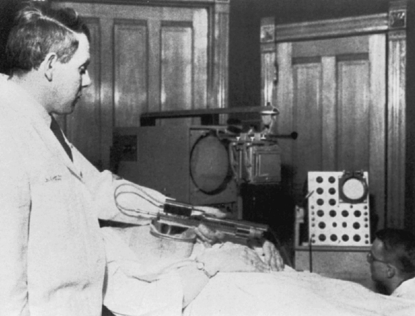
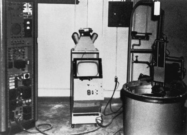
A, George D. Ludwig conducted A-mode experiments for the U.S. Navy to detect the presence of gallstones and other foreign bodies embedded in animal tissues. One of his primary concerns was establishing the physical and physiologic standards of the velocity of ultrasonic transmission through gallstones versus tissue. B, Scanning apparatus used by Ludwig to investigate sound transmission and the acoustic properties of tissue.
John A. Wild (left) applying a B-mode scanner to a patient’s breast, while John M. Reid operates the system controls (lower right).
A, The transducer assembly is mounted on the wooden track running along the outside edge of the tank. The actual transducer was submerged within the water-filled tank. B, Horizontal ultrasonic scan of a human leg produced by the cattle-tank scanner.
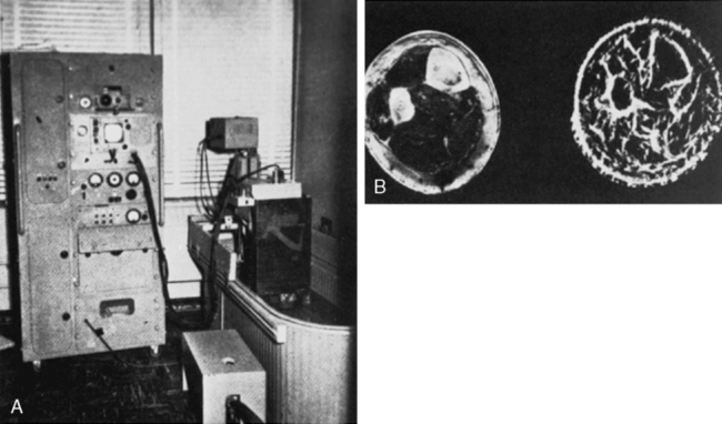
The transducer is assembly mounted on a ring encircling the tank. This arrangement provided a complete 360-degree scan-path around the immersed patient. The transducer could be raised and lowered to a desired level within the tank. A second motor moved the transducer in a 4-inch back-and-forth-sectoring motion. Left, The tall console on the left contained the electronic system; center, the display console; right, the gun turret and transducer carriage assembly.
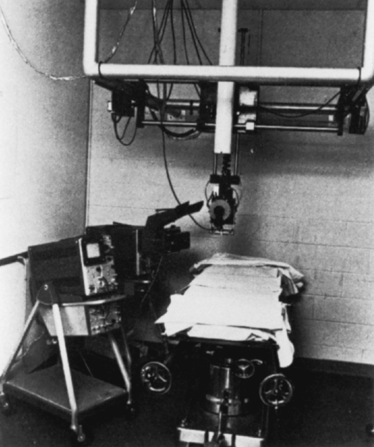
It was constructed by consulting engineers William Wright and Ed Meyer for Joseph Holmes. The system required one individual to manually guide the ceiling-mounted transducer carriage across the patient’s body, as the transducer mechanically sectored 30 degrees to either side of the perpendicular. A second individual operated the controls and the camera (left). This system was used during the 1960s to develop many of the scanning protocols still practiced today.
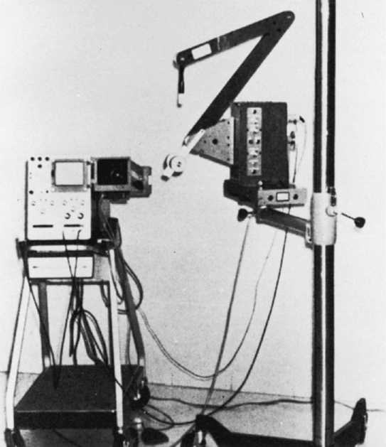
Engineers William Wright and Ed Meyer developed this portable compound contact scanner after forming their own company, Physionics Engineering. Freed from the ceiling mount and positioned upon a wheeled tripod, the transducer mechanism, a three-jointed scanning arm, and a mechanical and electronic “box” could be moved along a calibrated metal track. Horizontal or longitudinal positioning of the “arm” permitted scans in two planes. A single operator could now operate scanner, controls, and camera.
Left, A cross-sectional scan of the fetal head outlined in B-mode presentation. Right, An A-mode presentation of the midline echo. This scan (c. 1964) is thought to have been produced with the Diasonograph.
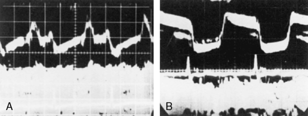
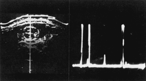
Carl Hellmuth Hertz and Inge Edler developed an ultrasonic technique to display graphic cardiac motion. Reflected echo information appeared as a bright dot moving along the screen as the structure being imaged moved or shifted position. A special camera and continuous moving film displayed the wave form motion of the echo dot reflected from the intracardiac structures. A, Normal mitral valve patterns. B, An abnormal, flat-topped stenotic mitral valve pattern.![]()
Stay updated, free articles. Join our Telegram channel

Full access? Get Clinical Tree


Radiology Key
Fastest Radiology Insight Engine