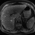
Fig. 1.1
Superficial spreading melanoma
1.2 Biopsy of the Lesion
Any clinically changing lesion should be further assessed with a biopsy. The recommended method is a complete excision biopsy, with direct closure, [4, 5, 12]. This allows full pathological assessment of the lesion confirming the diagnosis and providing prognostic information (Breslow thickness, presence of ulceration and mitotic rate) [5]. Wider margins are not recommended as this may interfere with subsequent lymphatic mapping [4, 13].
Partial biopsies, core, superficial or deep shave or incision biopsies may potentially underestimate the lesion leading to inadequate treatment. Difficult lesions such as Spitz tumours, nevoid or desmoplastic melanomas require the entire lesion to be excised to allow the most accurate pathological evaluation of the lesion [5].
Extremity biopsies should be aligned vertically along the limb to minimize the need for skin grafts or flaps [5]. Once the diagnosis has been made, definitive surgery can be undertaken.
1.3 Classification
There are four main types of primary melanoma.
1.3.1 Superficial Spreading Melanoma
It is the most common type of melanoma in Caucasians (see Fig. 1.1). The mean age of diagnosis is 51 years and comprises 40–60% of all melanomas [5, 8]. It frequently arises in a pre-existing naevus. It is often described as a slowly expanding variably pigmented lesion, before a period of rapid clinical change, often just before it is diagnosed [5, 8, 11]. It can occur at any site on the body but more frequently seen on the back in men and lower limbs in women.
1.3.2 Nodular Melanoma
It affects older persons with a mean age of 56 years, men more frequently than women. The overall incidenceis is 10–35% with a higher percentage of thick melanomas [5, 8]. It may develop in normal skin or a pre-existing naevus. They are often described as a rapidly changing lesion and can vary from a pink through to being darkly pigmented. If it lacks pigment it can resemble a vascular lesion. This makes the diagnosis difficult and can cause in a delay in presentation [5].
1.3.3 Lentigo Maligna Melanoma (LMM)
This melanoma classically develops on chronically sun-damaged skin in the elderly usually on the more sun-exposed areas (face and hands). It comprises 10–15% of melanomas [8]. This lesion usually develops from a precursor lesion called lentigo maligna or Hutchinson’s melanotic freckle (HMF). There is a 5% lifetime risk of HMF developing into the invasive melanoma without treatment [5, 8]. This is often associated with a desmoplastic component. Any nodule in an area of HMF should be investigated for LMM. The recurrence rate following standard excision is 8–20% [5, 8, 14–17].
1.3.4 Acrolentiginous Melanoma (ALM)
This type of melanoma develops in the acral skin of the palms and soles (see Fig. 1.2). It represents <5% of melanomas in countries with high rates of melanoma [8]. It is the most common type of melanoma in Blacks and Asian populations, with a mean age of diagnosis of 67 years. It is often light coloured or pink and relatively flat and often diagnosed late [5, 18].

Fig. 1.2
Acral melanoma
1.4 Classification/Staging
The eighth addition of the AJCC staging system is due to take effect in 2018 (Table 1.1). Although it is similar to the seventh addition, some changes have been made to the TNM classification. T0- is used when there is no evidence of primary tumour. TX- is used when Breslow thickness cannot be determined. Breslow thickne ss is now measured to the nearest 0.1 mm and not to two decimal points. T1 melanomas are further subcategorised by tumour thickness at a threshold of 0.8 mm. T1a- Is a nonulcerated melanoma <0.8mm. T1b- is between 0.8-1.0mm regardless of ulceration or ulcerated and <0.8mm. Mitotic rate is no longer used in the T category, but remains a major determinant of prognosis across all T categories. Regional lymph nodes and now defined as “clinically occult” or “ clinically detected” rather than microscopic or macroscopic nodal burden. Non-nodal regional disease such as microsatellites, satellites and in-transit metastases and now classified according to the number of involved lymph nodes. This is written as N1c, N2c or N3c. Serum lactate dehydrogenase is now classified as “0” when not elevated and “1” when elevated. This classification is attached to each M subgroup and is written in the format M1a(1). Central nervous system disease is classified as a separate new category of M1d. This is regardless of the involvement of other sites with metastatic disease. The AJCC prognostic stage groups are very similar to the seventh addition, however there is subtle differences between clinical TNM (cTNM) and pathological TNM (pTNM) (Table 1.2) [19, 21].
Table 1.1
TNM staging for melanoma based on the AJCC 8th Edition staging sytem (adapted from Amin MB et al. 2017 [22])
T stage | Tumour thickness (mm) |
|---|---|
Tx | Thickness unable to be assessed |
T0 | No evidence of primary tumour |
Tis | Melanoma in situ |
T1 | ≤1.0 mm |
T1a | <0.8 mm without ulceration |
T1b | <0.8 mm with ulceration or 0.8–1.0 mm without ulceration |
T2 | >1.0–2.0 mm T2a—without ulceration T2b—with ulceration |
T3 | >2.0–4.0 mm T3a—without ulceration, T3b—with ulceration |
T4 | >4.0 mm T4a—without ulceration T4b—with ulceration |
N stage | Number of involved nodes |
N0 | No nodes detected |
N1 | 1 node involved or In-transit, satellite and/or micro satellite metastases with no tumour involved nodes |
N1a | Clinically occult node & no in-transit/satellite/microsatellites |
N1b | Clinically detected node & no in-transit/satellite/microsatellites |
N1c | No regional node disease and in-transit/satellite/microsatellite metastases
Related posts:Stay updated, free articles. Join our Telegram channel
Full access? Get Clinical Tree
 Get Clinical Tree app for offline access

|