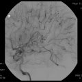
Benign meningioma (WHO grade I)
Any predominant histology other than clear cell, chordoid, papillary, or rhabdoid
Lacks criteria of atypical, brain invasive, and anaplastic meningioma
Atypical meningioma (WHO grade II) (any of two major criteria)
Mitotic index ≥4 mitoses/10 high-powered fields (HPF)
At least three of the following five parameters:
• Sheeting architecture (two dimensional sheets with loss of whorls and fascicles)
• Small cell formation (clusters of lymphocyte-like cells with high N/C ratio)
• Hypercellularity
• Macronucleoli
• Spontaneous necrosis (e.g., not induced by embolization or radiation)
Brain-invasive meningioma (WHO grade II)
Tongue-like protrustions of meningioma into adjacent brain parenchyma
Meningioma containing islands of entrapped GFAP-positive parenchyma
Anaplastic (malignant) meningioma (WHO grade III) (either of two criteria)
Mitotic index ≥20 mitoses/10 HPF
Frank anaplasia (sarcoma-, carcinoma-, or melanoma-like histology)
With appropriate treatment, approximately 80 % of benign (WHO grade I) or image-diagnosed meningiomas remain progression-free at 10 years (Fig. 22.1). In contrast, only about 35 % of atypical (WHO grade II) patients remain disease-free at 10 years even with definitive therapy [37, 39, 43]. Anaplastic (WHO grade III) meningiomas have a mean overall survival of less than 2–3 years and often progress quickly through serial treatments [39].


Fig. 22.1
Progression-free survival reported in 35 selected studies that included fractionated external beam radiation therapy by year of publication. Please note that technological advances in imaging, treatment planning and delivery primarily after the year 2000 resulted in superior and more consistent outcomes in both cohorts of patients treated with combined therapy (STR + RT) and RT alone. Advancements in surgical techniques over the same time period did not yield similar improvements in outcomes
External Beam Radiation Therapy: Benign Meningiomas
Benign Meningiomas
Although the majority of meningiomas are benign (WHO grade I) and slow-growing, they may become large enough over time to cause devastating neurologic deficits and morbidity if left untreated. Therefore, therapeutic intervention is often needed for most diagnosed tumors. In clinical practice, surgery has been the treatment of choice for most meningiomas. Resection enables histologic diagnosis, relieves mass effect and associated symptoms, alleviates tumor-induced seizures, and achieves high local control rates. However, not all tumors can be completely resected without considerable morbidity. The likelihood of safe and complete resection depends, in part, upon site and surgical accessibility, and is frequently precluded by involvement of the cranial nerves, arteries, or by venous sinus invasion. Gross total resection (GTR) is often not attempted in the cavernous sinus, clivus, cerebello-pontine angle, sellar region, or optic nerve sheath. This is significant since in his seminal publication in 1957, Simpson correlated the extent of tumor resection, associated dural attachments, and any hyperostotic bone to local recurrence risk, and defined 5 grades of resection, so-called Simpson grades (Table 22.2) [45]. The surgical resection extent has been consistently identified as an important prognostic feature [7, 12, 46, 47], and majority of centers continue to use Simpson’s criteria.
Table 22.2
Simpson grades of resection based on 265 patients
Resection grade | Definition | Recurrence (%) |
|---|---|---|
1 | GTR of tumor, dural attachments and abnormal bone | 9 |
2 | GTR of tumor, coagulation of dural attachments | 19 |
3 | GTR of tumor without resection or coagulation of dural attachments or extradural extensions (e.g., invaded or hyperostotic bone) | 29 |
4 | Partial resection of tumor | 44 |
5 | Simple decompression (biopsy) | – |
More recently Sughrue et al. challenged the relevance of Simpson’s classification in the modern era. With 373 WHO grade I meningioma patients followed for a median of 3.7 years, they found no significant difference in 5-year progression-free survival (PFS) following Simpson grade I–IV resections, with respective 5-year PFS rates of 95, 85, 88, and 81 % (p = ns) [48]. Similar findings were previously reported by Condra et al. [46] and more recently by Oya et al. [49]. Even though these studies did not identify a difference in local control after Simpson grade I–III resections, they did generally reveal shorter PFS following Simpson grade IV surgery [46, 49]. To directly address the predictive value of resection extent, Hasseleid et al. [50] reviewed 391 patients with convexity meningiomas and were able to identify significant outcome differences between Simpson grade I, grades II + III, and grade IV + V resections, continuing to support the relevance and applicability of Simpson’s criteria. Therefore, gross tumor resection (GTR, Simpson I–III) remains the objective of meningioma surgery, and is achievable in approximately one-half to two-thirds of patients [47, 51], depending on the tumor location [52]. For these situations, where complete and safe tumor removal is not possible, an effective strategy is STR followed by postoperative radiation therapy or radiation therapy alone.
For benign meningioma, GTR is considered as definitive treatment, but with extended follow-up, recurrences develop in approximately 5–30 % of patients (Table 22.3 and Fig. 22.1). For example, among 145 patients in whom GTR was achieved, Mirimanoff et al. [51] reported 5-, 10-, and 15-year recurrence rates of 7 %, 20 %, and 32 % and second operation rates of 6 %, 15 %, and 20 %, respectively. These rates were confirmed by a large series from the Mayo Clinic [12]. Condra et al. of the Unversity of florida, [46] reported the importance of complete excision but found no association between Simpson grade and local control or cause-specific survival, as long as GTR (Simpson grades 1, 2, and 3) was achieved. For patients treated with surgery alone, GTR resulted in 5-, 10-, and 15-year actuarial recurrence rates of 7 %, 20 %, and 24 %, respectively [46]. In a more recent report by Soyuer et al., the respective 5-, 10-, and 15-year rates were 23 % 39 %, and 60 %, the higher recurrence rates (Table 22.3) likely reflecting the more routine use and improved quality of neuroimaging [53].
Table 22.3
Selected studies of fractionated external beam radiation therapy for patients largely with WHO grade I or image-diagnosed (presumed WHO grade I) meningiomas
Author | Year | N | Technique | Progression-free survival | Clinical improvement (%) | Tumor shrinkage (%) | Late toxicity (%) | Time point (years) | |||
|---|---|---|---|---|---|---|---|---|---|---|---|
GTR (%) | STR (%) | STR + RT (%) | RT (%) | ||||||||
Adegbite et al. | 1983 | 114 | EBRT | 74 | 34 | 82 | 10 | ||||
Mirimanoff et al. | 1985 | 225 | EBRT | 80 | 45 | 10 | |||||
Barbaro et al. | 1987 | 135 | EBRT | 96 | 40 | 68 | 0 | Crude | |||
Taylor et al. | 1988 | 132 | EBRT | 77 | 18 | 82 | 10 | ||||
Glaholm et al. | 1990 | 117 | EBRT | 96 | 43 | 77 | 46 | 38 | 10 | ||
Miralbell et al. | 1992 | 115 | EBRT | 48 | 88 | 16 | 8 | ||||
Mahmood et al. | 1994 | 254 | EBRT | 98 | 62 | 5 | |||||
Goldsmith et al. | 1994 | 117 | EBRT | 77 | 3.6 | 10 | |||||
98a | |||||||||||
Peele et al. | 1996 | 86 | EBRT | 52 | 100 | 5 | Crude | ||||
Condra et al. | 1997 | 246 | EBRT | 80 | 40 | 87 | 24 | 10 | |||
Stafford et al. | 1998 | 581 | EBRT | 75 | 39 | 10 | |||||
Nutting et al. | 1999 | 82 | EBRT | 83 | 14 | 10 | |||||
Vendrely et al. | 1999 | 156 | EBRT | 79 | 59 | 29 | 8 | ||||
Pourel et al. | 2001 | 26 | EBRT | 76 | 2.2 | 8 | |||||
Dufour et al. | 2001 | 31 | EBRT | 93 | 71 | 29 | 3.2 | 10 | |||
Jalali et al. | 2002 | 41 | FSRT | 100 | 26.8 | 22 | 12.1 | 3 | |||
Uy et al. | 2002 | 40 | IMRT | 93 | 23 | 5 | 5 | ||||
Pirzkall et al. | 2003 | 20 | IMRT | 100 | 60 | 25 | 0 | 3 | |||
Soyuer et al. | 2004 | 92 | EBRT | 77 | 38 | 91 | 2.5 | 10 | |||
Selch et al. | 2004 | 45 | FSRT | 97 | 20 | 18 | 0 | 3 | |||
Milker-Zabel et al. | 2005 | 317 | IMRT | 89 | 42.9 | 23 | 0 | 10 | |||
Henzel et al. | 2006 | 224 | FSRT | 97 | 43.4 | 46 | 0 | 3 | |||
Milker-Zabel et al. | 2007 | 94 | IMRT | 94 | 39.8 | 20 | 4 | 4.4 | |||
Hamm et al. | 2008 | 183 | FSRT | 93 | 23.2 | 2.7 | 3 | ||||
Litre et al. | 2009 | 100 | FSRT | 94 | 94 | 50–81 | 9 | 0 | 5 | ||
Korah et al. | 2010 | 41 | FSRT | 94 | 3 | 5 | |||||
Metellus et al. | 2010 | 53 | FSRT | 94 | 94 | 58.5 | 30 | 1.9 | 10 | ||
Bria | 2011 | 60 | FSRT | 95 | 60 | 1 | |||||
96 | 3 | ||||||||||
Minniti | 2011 | 52 | FSRT | 93 | 20 | 23 | 5.5 | 5 | |||
Mahadevan | 2011 | 16 | FSRT | 100 | 19 | 4 | 2 | ||||
Morimoto | 2011 | 31 | FSRT | 87 | 1 | 5 | |||||
Onodera | 2011 | 27 | FSRT | 96.2 | 5.3 | ||||||
Ohba | 2011 | 281 | FSRT/SRS | 88.3 | 63.7 | 92.3 | 5 | ||||
96 | 99 | 5 | |||||||||
Tanzler | 2011 | 146 | EBRT/FSRT/SRS | 93 | 99 | 6.8 | 10 | ||||
Paulsen | 2012 | 109 | FSRT | 98 | 21 | 5 | 5 | ||||
Recurrence rates following STR alone are substantially higher (Table 22.3 and Fig. 22.1). For example, among 116 patients with STR, Stafford et al. [12] found recurrences in 39 % at 5 years and 61 % at 10 years. Condra et al. [46] documented local recurrences in 47 %, 60 %, and 70 % of patients with STR at 5, 10, and 15 years, respectively. Overall, it is estimated that 40–50 % of patients with STR develop local progression within 5 years, 60 % within 10 years, and at least 70 % within 15 years [46, 47]. In a recent study, Jensen et al. [54] reported that STR was associated with both poorer PFS and overall survival (OS). Despite these data, patients are frequently observed following STR. For example, in a Mayo Clinic series of 581 patients, 116 (20 %) of whom has STR, only 10 (9 %) received adjuvant radiation therapy [12].
For cases where GTR is not possible, radiation therapy (RT), whether fractionated external beam radiation therapy (EBRT) or stereotactic radiosurgery (SRS), is currently the only accepted nonsurgical treatment option. Historically, meningiomas were considered to be resistant to RT, and authors of many older retrospective studies reported infrequent radiographic regression [55]. Additionally, concerns about malignant degeneration were cited as a reason to refrain from either definitive or postoperative RT [29, 56]. Consequently, many patients with inoperable or subtotally resected meningiomas were observed. At present, malignant degeneration has not been explicitly linked to RT [32, 51, 57]; rather, it is believed to be related to the natural history of a subset of meningiomas [58, 59].
Most retrospective studies since the 1980s have demonstrated that, for benign meningioma, postoperative RT after STR improves local control (Table 22.3 and Fig. 22.1), and perhaps even survival [46, 60]. Improvement in local control, and frequently in tumor-related neurologic symptoms, was observed despite the fact that only about one-fourth to one-third of treated meningiomas exhibit tumor shrinkage (Table 22.3).
Radiotherapy can also be used as a primary treatment for meningiomas diagnosed radiographically or by biopsy. In one of the earliest reports, Glaholm et al. [29, 56] reported the outcome of 32 patients treated with primary RT without resection at the Royal Marsden Hospital; 47 % disease-free survival (DFS) was observed at 15 years. This was only modestly lower than a corresponding 56 % DFS in patients treated with STR + RT. More recent series demonstrate markedly improved local control rates and PFSs, >93 % after 5–10 years of follow-up (Table 22.3) [61–64]. These outcomes compare favorably to those observed after GTR for benign meningiomas (Fig. 22.1) and, to a certain degree, challenge the paradigm that surgery is the mainstay of meningioma management.
The discrepant outcomes of older studies (before 2000) versus more recent RT studies, whether in the postoperative or primary setting, may be attributed to the technical advancements in RT treatment planning and delivery techniques. This trend is clearly illustrated in Fig. 22.1 for PFS, and in Table 22.3 for RT-related late neurologic toxicity as well as neurologic improvement. These observations are more directly supported by the analysis of Goldsmith et al. [65] of 140 patients treated at the University of California, San Francisco to a median dose of 54 Gy in the postoperative setting where a PFS rate of 98 % was noted in those treated after 1980 (when computed tomography [CT] and/or magnetic resonance imaging [MRI] was used for planning therapy) compared to a rate of 77 % for patients treated before 1980 when advanced planning techniques were not available.
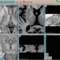
Newer treatment series also report the use of intensity-modulated RT (IMRT) as well as fractionated stereotactic RT (FSRT) (Table 22.3). IMRT techniques allow delivery of dose via multiple convergent beams (Fig. 22.2), each of which can be divided into zones of different intensities, so that the integral dose can tightly conform to the shape of the target. This can potentially minimize unnecessary irradiation of the surrounding normal tissues, including the critical anterior optic structures. FSRT is characterized by highly accurate patient immobilization setup, use of a stereotactic coordinate system, and sub-millimeter mechanical and beam tolerances. All modern RT techniques rely on the use of sophisticated imaging for treatment planning and verification. It is believed, but not yet definitively proven, that these newer treatment methods will continue to improve patient outcomes and reduce RT-related toxicity.


Fig. 22.2
A representative intensity-modulated radiation therapy (IMRT) plan for a right cavernous sinus meningioma (WHO grade I) in a 52 years old patient with slight right temporal field deficit thought to be related to disease encroachment on the right portion of the optic chiasm. GTV is shaded red; a dose of 54 Gy/30 fractions was prescribed to the 95 % isodose line to minimize the risk of injury to the anterior optic structures. Note: Maximal point dose for the plan (56.38 Gy) is located within the tumor volume, and the maximum dose to the chiasm is <54 Gy
It is worth noting that serious side effects of modern methods of treatment planning and delivery are already quite uncommon. For example, Debus et al. [66] reported on 189 patients treated with FSRT for large skull base meningiomas, using median daily fractions of 1.8 Gy to a mean dose of 56.8 Gy. With a nearly 3-year median follow-up, the rate of clinically significant neuro-toxicity (grade 3) was 2.2 % (four patients), and 1.7 % (three patients) in the absence of a preexisting neurologic deficit. The neurologic deficits reported in the study included reduced vision, a new visual-field loss, and trigeminal neuropathy. Goldsmith et al. [65] noted complications in 3.6 % of 140 patients treated with RT after STR from 1967 to 1990. The observed RT-related toxicity in this study included retinopathy, optic neuropathy, and cerebral necrosis. Further analysis of the data by Goldsmith et al. [67] permitted construction of a model to predict that optic nerve tolerance is 890 optic ret, equivalent to 54 Gy/30 fractions. This threshold dose is supported by only rare observations of optic nerve injury with doses lower than 54 Gy, particularly with fractional doses ≤2.0 Gy [56, 68, 69]. Additionally, Uy et al. [70] noted no anterior optic pathway injury with a median dose of 50.4 Gy and fractions of 1.7–2.0 Gy. Shrieve et al. [71] confirmed these findings as well as the benefit of fractional doses at or lower than approximately 2.1 Gy.
Other cranial neuropathies (non-optic) are uncommon with modern treatment techniques but have been reported in older series. For example, Selch et al. [72] found no RT-related neuropathy in 45 patients treated for cavernous sinus meningiomas to median dose of 50.4 Gy (1.8 Gy/fraction). Similar findings were noted earlier by Urie et al. [73]. Brain or brainstem injury, including necrosis, is quite uncommon in the modern era with conventional doses and techniques. Similarly, edema is generally not observed following fractionated RT, unlike single-session SRS treatment. For this reason, fractionated RT may be preferred over SRS for tumors in locations where edema is likely to develop and to significant morbidity (e.g., parasagittal, cerebello-pontine angle, and/or large meningiomas).
Optic Nerve Sheath Meningiomas
The management of optic nerve sheath meningiomas (ONSMs) is almost exclusively nonsurgical. These rare tumors comprise <2 % of all meningiomas and arise from the meningeal lining of the optic nerves [74, 75]. They generally grow very slowly within the subarachnoid space and the confines of the optic canal, eventually compressing the optic nerve itself and/or its vasculature. Patients usually present with progressive vision loss, although some exhibit rapid loss of vision. In a minority of patients, ONSMs are found incidentally on imaging. Less common complaints include visual-field defects, loss/alterations of color vision, and orbital pain.
Resection of ONSMs while keeping optic nerve in place has been tried [76–80], but carries an unacceptably high risk of visual complications and local recurrence. Resection commonly impairs the blood supply and results in blindness, even though the nerve is left grossly intact. For these reasons, fractionated RT has become the centerpiece of ONSM management. In one of the largest series published, Turbin et al. [80] reported on 64 patients with ONSMs and found that fractionated RT alone provided more favorable outcomes than observation, surgery alone, or a combination of surgery + RT. Patients in the RT only group were the only ones that did not demonstrate a statistically significant decline in visual acuity after a mean follow-up of 11.5 years (range, 4.75–23 years); all other groups demonstrated a significant decrement in visual acuity, including complete visual loss. Irradiated patients received 40–55 Gy of fractionated RT (1.8–2.0 Gy/fraction) over approximately 5 weeks. In a study from Japan, Narayan et al. [81] evaluated 14 patients with ONSMs who were treated with conformal, fractionated RT to 50.4–56 Gy. After a median follow-up of 4.1 years, 86 % of the treated patients had either improved or stable visual acuity. These studies and six additional studies with similar results have established highly conformal, fractionated RT as the first-choice treatment modality for patients with ONSMs. In aggregate, the local control is excellent (95 %); early visual improvement (<3 months from the completion of RT) is attained in about half of the patients (54.7 %); and complications of therapy are relatively uncommon [74, 82]. It is our policy to treat symptomatic ONSM patients with highly conformal, fractionated RT to doses of 50.4–54 Gy using daily fractions of 1.8 Gy; however, there is no uniform agreement as to whether patients with stable ONSMs and stable vision should be approached with observation or early RT. Patients with bilateral disease are generally treated whereas those with unilateral tumors are observed provided that they can be expected to comply with serial visual field testing, and regular clinical and imaging follow-up.
Stay updated, free articles. Join our Telegram channel

Full access? Get Clinical Tree


