(a)
The origin of the celiac artery (CA) is at the level of the 12th thoracic vertebra and behind the median arcuate ligament. Following a short anterior course, the CA commonly divides into three main branches: the common hepatic artery, the left gastric artery, and the splenic artery. These arteries supply the foregut.
(b)
The superior mesenteric artery (SMA) originates at the level of the first lumbar vertebra. It lies anterior to the left renal vein and posterior to the body of the pancreas. The SMA supplies the mid-gut (small intestine from the second part of the duodenum to the midtranverse colon), and sections of the pancreas.
(c)
The inferior mesenteric artery (IMA) arises from the aorta approximately 5 cm above the aortic bifurcation (level of the third lumbar vertebra) and supplies the left hemicolon and the proximal part of the rectum.
2.
Numerous arterial interconnections exist between the three mesenteric main branches and form an abundant collateral circulation. The gastroduodenal and pancreaticoduodenal arteries form a collateral network between the CA and the SMA. The middle colic artery also links the SMA to the IMA via the left colic artery. The arterial complex of Drummond (the marginal artery in the vasa recta of the colon and the central anastomotic artery or loop of Riolan) and the arc of Riolan are interconnections between the superior and inferior mesenteric circulation.
2 Disease Definition
Mesenteric ischemia: significant stenosis or occlusions of at least two mesenteric arteries or single-vessel obstruction without sufficient collateral circulation leading to symptoms like food fear, loss of weight, and postprandial pain.
3 Disease Distribution
Unselected autopsy studies have estimated the prevalence of mesenteric atherosclerosis in at least one mesenteric artery to be up to 6–10%. The exact incidence of chronic mesenteric ischemia (CMI) has been estimated to be 1 in 100,000 of the general population per year. CMI accounts for 0.1% of all hospital admissions and 1% of the admissions in patients with acute abdominal pain. The average age of patients with CMI diagnosed is 60 years, whereas acute mesenteric ischemia (AMI) is often determined in older patients.
4 Diagnosis: Clinical and Laboratory
CMI is an uncommon disease. The symptoms may be nonspecific and vary from the classical triad: postprandial abdominal pain leading to food fear, weight loss, and upper abdominal bruit to nonspecific abdominal pain and intermittent diarrhea. Laboratory findings will not be anomalous in patients with CMI.
5 Diagnostic Imaging
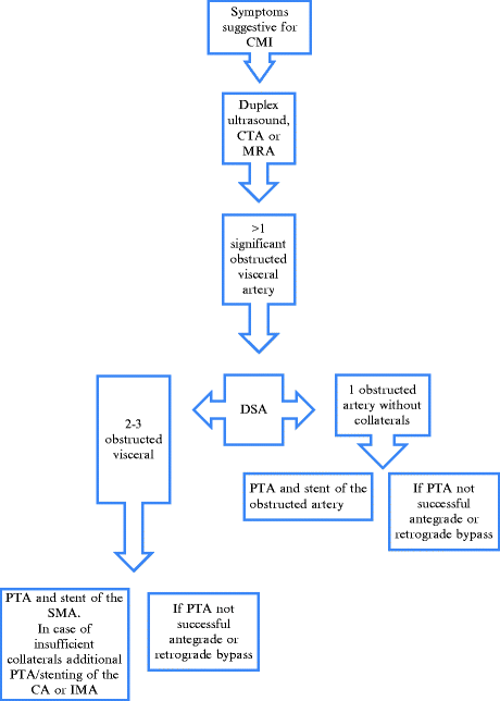
A critical point in treating patients with chronic CMI—both open and with endovascular means—is adequate diagnostic imaging. Imaging studies to identify the presence of mesenteric artery obstructive disease include Duplex ultrasound (DUS), magnetic resonance angiography (MRA), or computed tomography angiography (CTA). CMI is suspected in patients with clinical suspicion of mesenteric ischemia and at least two mesenteric arteries demonstrating a hemodynamically significant stenosis or occlusion. Single-vessel visceral obstruction without sufficient collateral circulation may also lead to symptomatic CMI although this is uncommon.
1.
DUS is a good noninvasive modality for obstructive disease of the CA and SMA. Sensitivity, specificity, and predictive values are all >80%. A normal duplex examination excludes the diagnosis of CMI in most of the patients. Drawbacks of DUS are the fact that the IMA may be difficult to visualize and the fact that the side branches and collaterals cannot be determined.
2.
MRA combines excellent sensitivity, specificity, and predictive values with limited contrast load. It allows the determination of obstructive disease in all main visceral arteries and major collaterals. However, high-resolution MRA is not universally available, technician dependent, and some patients are excluded (metal artifacts, cardiac prosthetic valves, claustrophobia).
3.
Multidetector CTA has replaced diagnostic catheter-based contrast arteriography as the imaging study of choice during last years. Besides its accuracy of detecting visceral artery obstruction (including side branches and collaterals) it will exclude other potential intra-abdominal processes in patients with abdominal complaints. CTA is sufficient to plan open visceral reconstructive surgery and useful as follow-up modality of endovascular and open revascularization procedures.
4.
Diagnostic catheter based digital subtraction angiography (DSA) has been replaced by noninvasive CTA and/or DUS. In most of the patients with indication for DSA, diagnostic imaging will be combined with direct endovascular treatment in the same setting.
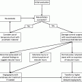
6 Management Paradigm and Indications (Table 21.1)
Patients presenting with longstanding symptoms suggestive of CMI and proven visceral obstructive disease on vascular imaging must be discussed in a multidisciplinary workgroup consisting of interventional radiologists, gastroenterologists, and vascular surgeons. Other diagnoses have to be excluded by abdominal CT and (on indication) gastroscopy or colonoscopy, or both. Gastric and jejunal exercise tonometry can be used as a functional test (similar to the treadmill test in patients with peripheral arterial disease) and is used by the authors on indication. In patients with high suspicion for CMI the diagnosis can be obtained by digital subtraction angiography (DSA); endovascular treatment is performed in the same session.
7 List of the Open Operative Choices
Related posts:

Stay updated, free articles. Join our Telegram channel

Full access? Get Clinical Tree


