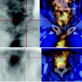
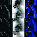
Fig. 46.1
PET-CT: in the upper quadrants of the left breast a rough solid, irregular lesion, with modest increases in glucose metabolism is detected. Intense consumption of FDG at the level of a backbone metamer and the sternum due to post-chemotherapy rebound is also found

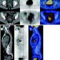
Fig. 46.2
CT-PET: the left iliac bone presents a lytic roundish area with interruption of the cortical profile; this element is suggestive of a metastatic lesion although focal carbohydrate consumption is not shown. The dissociation between metabolism and morphology of the lytic lesion is determined by the response to neoadjuvant chemotherapy. This element is confirmed by the fusion image
46.5 Key Points
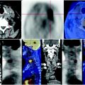
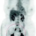
The PET scan shows the good metabolic response of the lesions at the left axilla and ipsilateral iliac bone, even if the primary tumor has moderate persistence of residual biological activity.
Stay updated, free articles. Join our Telegram channel

Full access? Get Clinical Tree


