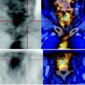
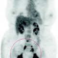
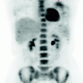
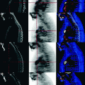
Fig. 41.1
MIP reconstruction: there are no focal lesions with a high glucose uptake, in particular the mediastinal mass shows metabolic activity comparable to that of the liver
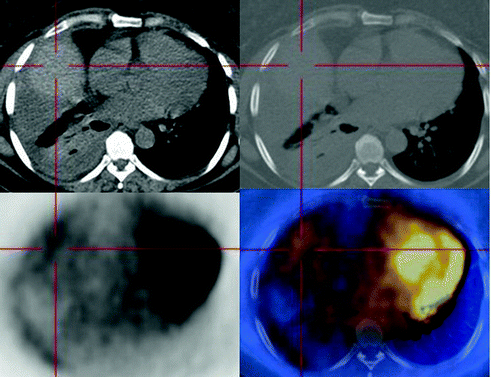
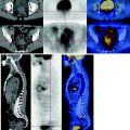
Fig. 41.2


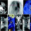
The PET-CT scan shows opacification of the pulmonary medium and basal fields average with right pleural effusion, characterized by irregular density for the presence of areas of atelectasis determined by recent FNAB, with reduced expansion of the right hemithorax and hemidiaphragm lifted upwards (a). The recent fine-needle aspiration path is best evident in the sagittal reconstruction of the CT-PET, documenting the increased density of the soft tissues of the anterior wall of the ipsilateral hemithorax (b). The lesion carbohydrate consumption is low, comparable to that of the mediastinal background
Stay updated, free articles. Join our Telegram channel

Full access? Get Clinical Tree


