CHAPTER 3 Middle Ear and Mastoid Air Cells
Embryology
Pharyngeal pouches develop between the branchial arches (the first pouch is found between the first and second branchial arches) in very early embryonic life. The endoderm of the pharyngeal pouches and the ectoderm of the branchial grooves contact each other to form the branchial membranes separating the pharyngeal pouches and the branchial grooves. The epithelium of the first branchial groove briefly touches the endoderm of the first pharyngeal pouch at the site of future tympanic membrane.
The first pharyngeal pouch expands to form a tubotympanic recess. This tubotympanic recess gives rise to the tympanic cavity, the mastoid antrum, and their epithelial lining. Connection between the tubotympanic recess and the pharynx elongates to form the auditory tube. The middle ear ossicles are derived from the first and the second branchial arches. The first branchial arch contributes to bodies of malleus and incus and the second arch forms the crura of the stapes, the lenticular process and long process of the incus, and the manubrium of the malleus.
Anatomy of the Middle Ear
The middle ear is an irregular, air-filled space within the temporal bone. It communicates with the nasopharynx through the auditory tube. It contains a chain of movable bones, which connect its lateral to its medial wall, and serve to convey the vibrations communicated to the tympanic membrane across the cavity to the internal ear. The middle ear can be divided into three parts (Fig. 3-1): the mesotympanum or the tympanic cavity proper that lies opposite the tympanic membrane; the epitympanum (or the attic) that lies cranial to the tympanic membrane; and the hypotympanum that forms a variable inferior extension of the middle ear cavity below the level of the tympanic membrane.
The tympanic membrane (TM) consists of three layers. The outer layer is continuous with the squamous epithelium of the external auditory canal (EAC). The central layer consists of fibrous connective tissue and is derived from the mesoderm. The inner mucous membrane is continuous with the lining of the inner ear. The circumference of the TM is thickened and forms a fibrocartilaginous ring that is fixed to the tympanic sulcus at the inner end of the EAC. This sulcus is deficient superiorly at the notch of Rivinus. Two bands, the anterior and posterior malleolar folds, take origin from the edges of the notch of Rivinus and extend inferiorly to the lateral process of the malleus. The smaller, triangular part of the TM situated above these folds is lax and thin, and is termed pars flaccida. The manubrium of the malleus is firmly attached to the medial surface of the membrane at a depression called the umbo. The remaining TM is taut and is referred to as the pars tensa. The Prussak space is an important space that is delineated laterally by the pars flaccida, superiorly by that lateral malleolar ligament, inferiorly by the short process of malleus, and medially by the malleolar neck. The epitympanum is bounded laterally by the scutum, derived from the squamous portion of the temporal bone(Fig. 3-2).
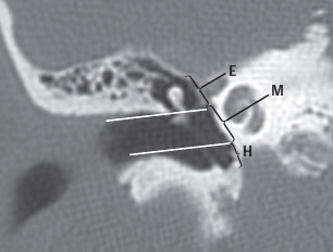
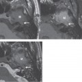
Figure 3-1 Coronal localizer that helps define the location of the epitympanum (E), mesotympanum (M), and hypotympanum (H).
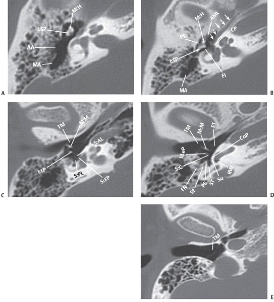
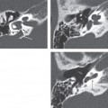
Figure 3-2
Related posts:

Stay updated, free articles. Join our Telegram channel

Full access? Get Clinical Tree


