Fig. 10.1
Transesophageal echocardiography (TEE) four-chamber view showing ruptured chordae tendineae of the posterior leaflet. The ruptured chordae (arrow) are seen as a freely moving structure from the left ventricle to the left atrium during the cardiac cycle. LA left atrium, LV left ventricle, RV right ventricle
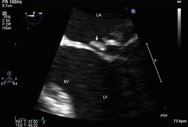
Fig. 10.2
A TEE zoom view of the mitral valve demonstrating flail of the posterior mitral leaflet with ruptured chordae (arrow). LA left atrium, LV left ventricle, RV right ventricle
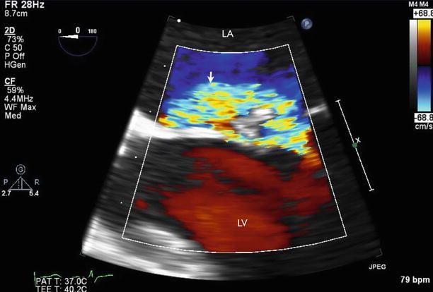
Fig. 10.3
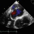
TEE zoom view of the mitral valve with color flow Doppler imaging demonstrating anteriorly directed mitral regurgitation jet (arrow). LA left atrium, LV left ventricle
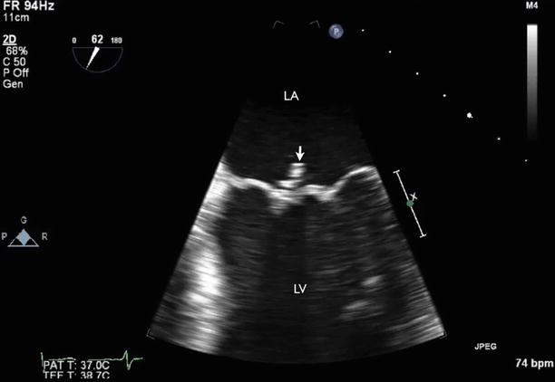
Fig. 10.4
TEE at 60° showed ruptured chordae with papillary muscle head moving freely into the left atrium (arrow). LA left atrium, LV left ventricle
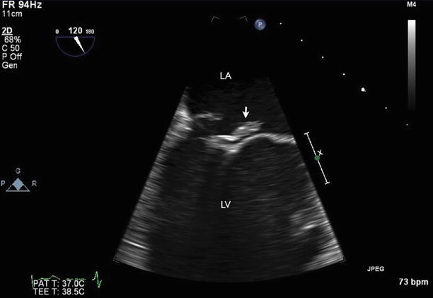
Fig. 10.5
TEE at 120° showed ruptured chordae with papillary muscle head moving freely into the left atrium (arrow). LA left atrium, LV left ventricle
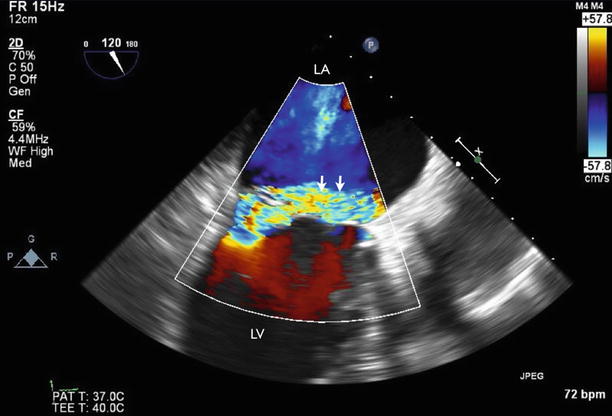
Fig. 10.6
TEE at 120° with color flow Doppler imaging demonstrated anteriorly directed mitral regurgitation jet (arrows). LA left atrium, LV left ventricle
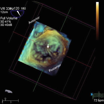
Fig. 10.7
Three-dimensional (3D) reconstruction imaging of the mitral valve (surgical view) showed a flail P3 segment of the mitral valve with ruptured papillary muscle (arrow)
Video 10.1 Transesophageal echocardiography (TEE) four-chamber view showed ruptured chordae tendineae of the posterior leaflet. The ruptured chordae are seen as a freely moving structure from the left ventricle to the left atrium during the cardiac cycle (AVI 5505 kb)
Video 10.2 A TEE zoom view of the mitral valve demonstrating flail of the posterior mitral leaflet with ruptured chordae (AVI 12633 kb)
Video 10.3 TEE zoom view of the mitral valve with color flow Doppler imaging demonstrating anteriorly directed mitral regurgitation jet (AVI 3203 kb)
Video 10.4 TEE at 60° showed ruptured chordae with papillary muscle head moving freely into the left atrium (AVI 9826 kb)
Video 10.5 TEE at 120° showed ruptured chordae with papillary muscle head moving freely into the left atrium (AVI 10014 kb)
Video 10.6 TEE at 120° with color flow Doppler imaging demonstrating anteriorly directed mitral regurgitation jet (AVI 1972 kb)
Video 10.7 Three-dimensional (3D) reconstruction imaging of the mitral valve (surgical view) showing flail P3 segment of the mitral valve with ruptured papillary muscle (AVI 2454 kb)
10.1.1 Learning Points
Papillary muscle rupture (PMR) is a catastrophic mechanical complication of acute myocardial infarction (AMI). Patients with PMR typically present 2–7 days after AMI with acute pulmonary edema and are often in cardiogenic shock. The absence of new heart murmur after AMI does not exclude the diagnosis. PMR is far more common in patients with inferior wall infarction because the posteromedial papillary muscle has a single blood supply via the posterior descending coronary artery, whereas the anterolateral papillary muscle has a dual blood supply from both the left anterior descending and the left circumflex coronary arteries. The ruptured head or heads are seen to be attached to the leaflets by the mitral chordae. These can mimic vegetations, but the clinical presentation, the chordal attachment to the leaflets, and the usual attachment to both leaflets help to distinguish them from vegetations.
Stay updated, free articles. Join our Telegram channel

Full access? Get Clinical Tree


