Mock-Board Review Questions
To test your active recall without spending hours making materials manually, tools like Quizgecko can transform your existing lecture notes into custom practice quizzes and flashcards in seconds.
QUESTIONS
Question 1
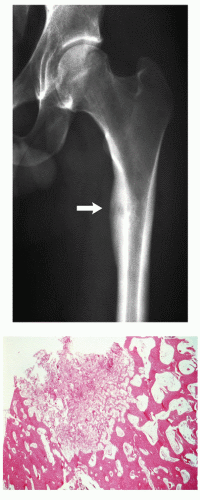
A 22-year-old man presented with nocturnal pain in the left groin. The anteroposterior radiograph of the left hip and histopathologic section of the lesion removed from the medial cortex of the femur are shown below. What is your diagnosis?
 |
A) Osteoid osteoma.
B) Chondroblastoma.
C) Osteofibrous dysplasia.
D) Osteosarcoma.
E) Ewing sarcoma.
View Answer
Answer to question 1: A
The radiograph shows a radiolucent lesion (arrow) within the medial femoral cortex exhibiting narrow zone of transition and surrounded by sclerotic area. There is associated well-organized periosteal reaction. Histopathologic section shows sharply circumscribed lesion containing bone trabeculae within vascularized connective tissue, lined by the plump osteoblasts and surrounded by sclerotic bone. Both, imaging characteristics and histopathologic features, are consistent with osteoid osteoma. (B) Chondroblastoma usually is present in the immature skeleton and is invariably located in the epiphysis. Histopathology shows chondroid matrix and not osteoblasts. (C) Osteofibrous dysplasia predominantly occurs in childhood (more than 60% of the patients are younger than 12 years old) and affects the cortex of the tibia (not femur). Histopathology shows characteristic immature woven bone trabeculae with peripherally displayed lamellar bone, lined by osteoblasts (“dressed trabeculae”). (D) Osteosarcoma and (E) Ewing sarcoma are predominantly intramedullary tumors, exhibiting more aggressive imaging characteristics. Histopathology shows malignant cells.
Question 2
Association of Ollier disease and hemangiomas is known as:
A) von Recklinghausen disease of bone.
B) Li-Fraumeni syndrome.
C) Maffucci syndrome.
D) Hereditary multiple exostoses.
E) Gardner syndrome.
View Answer
Answer to question 2: C
Maffucci syndrome is characterized by multiple enchondromas (Ollier disease) and cavernous hemangiomas located in the skin and soft tissues. This syndrome carries a greater risk of chondrosarcoma as well as nonskeletal malignancies, particularly of the ovaries and brain. (A) von Recklinghausen disease of bone, also known as osteitis fibrosa cystica generalisata, refers to severe bone disorder of primary hyperparathyroidism, characterized by increased osteoblastic activity, osteoclastic resorption, peritrabecular fibrosis, and brown tumors. (B) Li-Fraumeni syndrome caused by inherited mutation of p53 is associated with greater risk of sarcomas (particularly osteosarcoma) and other malignancies, such as breast cancer, brain tumors, and leukemias. (D) Hereditary multiple exostoses, also known as diaphyseal aclasis, is an autosomal dominant disorder characterized by the presence of multiple osteochondromas, particularly at the knee, hip, ankle, and shoulder joints. (E) Gardner syndrome, an autosomal dominant disorder, consists of a combination of multiple osteomas, intestinal polyposis, skin fibromatosis, and epidermal sebaceous cysts.
Question 3
Which of the following is associated with high risk of osteosarcoma?
A) Germ-line mutation of p53.
B) Translocation t(X;18).
C) Translocation t(12;16).
D) 1p rearrangements.
E) Nonrandom translocation t(9;22).
View Answer
Answer to question 3: A
Mutation of p53 (Li-Fraumeni syndrome) is associated with greater risk of sarcomas, particularly 500-fold greater risk of osteosarcoma. (B) Translocation t(X;18) is characteristic of synovial sarcoma. (C) Translocation t(12;16) is characteristic of myxoid liposarcoma. (D) 1p rearrangements are characteristic for conventional chondrosarcoma. (E) Translocation t(9;22) is characteristic for myxoid chondrosarcoma.
Question 4
Which one of the following should NOT be included in the differential diagnosis of Ewing sarcoma?
A) Lymphoma.
B) Metastatic neuroblastoma.
C) Mesenchymal chondrosarcoma.
D) Leiomyosarcoma of bone.
E) Metastatic embryonal rhabdomyosarcoma.
View Answer
Answer to question 4: D
(A) Lymphoma, (B) metastatic neuroblastoma, (C) mesenchymal chondrosarcoma, and (E) embryonal rhabdomyosarcoma show similar morphologic features to Ewing sarcoma, all characterized by small round blue tumor cells. Leiomyosarcoma of bone, on the other hand, is composed of bundles of fascicles of spindle cells.
Question 5
Which one of the following osteosarcomas has a better prognosis?
A) Paget osteosarcoma.
B) Periosteal osteosarcoma.
C) Parosteal osteosarcoma.
D) Telangiectatic osteosarcoma.
E) Small cell osteosarcoma.
View Answer
Answer to question 5: C
Parosteal osteosarcoma is low-grade (grade 1) sarcoma. (B) Periosteal osteosarcoma is a grade 2, and (A) Paget osteosarcoma, (D) telangiectatic osteosarcoma, and (E) small cell osteosarcoma are high-grade (grade 3) sarcomas.
Question 6
Adamantinoma of long bones is associated with the following features EXCEPT one:
A) The most common histopathologic variants are basaloid, spindle, squamous, and tubular.
B) The epithelial component expresses positivity for cytokeratins 8 and 18.
C) Younger age of the patients is associated with better prognosis.
D) An osteofibrous dysplasia-like type is the least common histopathologic variant.
E) Pain at presentation is associated with less favorable prognosis.
View Answer
Answer to question 6: B
The epithelial component of adamantinoma expresses positivity for cytokeratins 14 and 19. CK8 and CK18 are expressed in epithelioid component of synovial sarcoma. The statements (A), (C), (D), and (E) are all correct.
Question 7
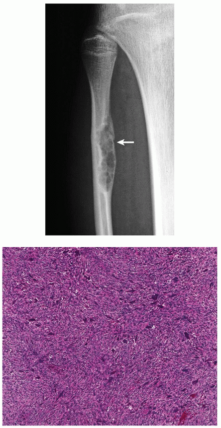
A 14-year-old girl injured her right knee in a fall. The radiograph demonstrated a lesion in the proximal diaphysis of fibula (arrow). An open biopsy was performed. Based on the radiographic presentation and histopathologic section, what is your diagnosis?
 |
A) Giant cell tumor (GCT)
B) Nonossifying fibroma (NOF)
C) Aneurysmal bone cyst (ABC)
D) Chondroblastoma
E) Giant cell-rich osteosarcoma
View Answer
Answer to question 7: B
The radiograph shows well-defined eccentric radiolucent lesion with sclerotic lobulated border, exhibiting no evidence of periosteal reaction (arrow), all imaging features of nonossifying fibroma. Histopathology shows fibrous stroma displaying a storiform arrangement, and several scattered giant cells, confirming the radiologic diagnosis. (A) Giant cell tumor almost always occurs in skeletally mature patients and extends into the articular end of bone. Generally, it does not exhibit a sclerotic border. Histopathology shows mononuclear cells intermixed with numerous uniformly distributed giant cells. (C) Aneurysmal bone cyst characteristically shows expansive features and well-organized periosteal reaction in the form of a buttress. Histopathology shows multiple blood-filled spaces alternating with more solid areas. (D) Chondroblastoma is an epiphyseal lesion, exhibiting chondroid calcifications. Histopathology shows nodules of fairly mature cartilaginous matrix surrounded by a highly cellular tissue composed of round and polygonal chondroblast-like cells, and matrix calcifications having a spatial arrangement that resembles the hexagonal configuration of chicken wire. (E) Giant cell-rich osteosarcoma exhibits aggressive imaging features with poorly defined borders showing wide zone of transition. Histopathology is similar to that of a giant cell tumor; however, tumor bone or tumor osteoid is present in the vicinity of malignant cells.
Question 8
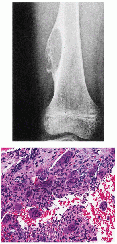
An 8-year-old boy presented with pain in his left lower thigh knee for past 3 months. Based on the submitted radiograph and histopathologic section, what is the most likely diagnosis?
 |
A) Nonossifying fibroma (NOF).
B) Aneurysmal bone cyst (ABC).
C) Simple bone cyst (SBC).
D) Giant cell tumor (GCT).
E) Telangiectatic osteosarcoma.
View Answer
Answer to question 8: B
The radiograph shows an eccentric radiolucent lesion with narrow zone of transition and geographic type of bone destruction, exhibiting a noninterrupted periosteal reaction in form of a buttress, all typical imaging features of aneurysmal bone cyst (ABC). Histopathology shows plump fibroblasts and giant cells aggregating around and within the blood-filled spaces, some of them showing characteristic feature of “jumping into the swimming pool”, confirming the radiologic diagnosis. (A) Nonossifying fibroma exhibits no expansive imaging features and lack of periosteal reaction. Histopathology shows predominantly bland monomorphous spindle cells admixed with histiocytic cells frequently arranged in a swirling storiform pattern and occasionally scattered giant cells. (C) Simple bone cyst shows almost invariably central location and lack of periosteal reaction, unless there is a pathologic fracture present. Histopathology shows walls of the cyst covered by a flattened single-cell lining. (D) Giant cell tumor, although occasionally may exhibit expansive imaging features, generally shows lack of sclerotic border and lack of buttress type of periosteal reaction, characteristic features of ABC. Histopathology shows mononuclear cells intermixed with numerous uniformly distributed giant cells. (E) Telangiectatic osteosarcoma, although occasionally similar in appearance to ABC, shows more aggressive imaging features associated with a soft-tissue mass. Interrupted periosteal reaction in form of lamellar, sunburst, or Codman triangle is commonly present. Histopathology shows malignant cells forming tumor bone or tumor osteoid.
Question 9
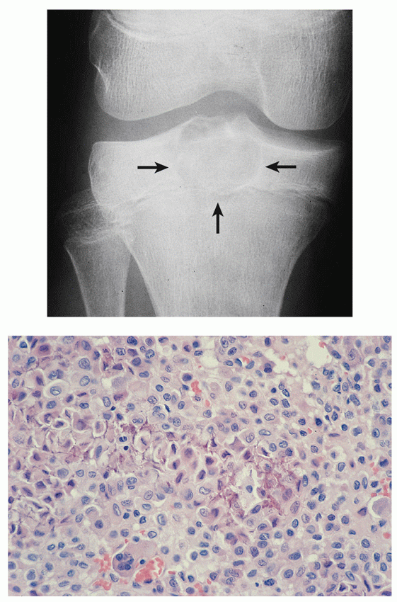
An 8-year-old boy presented with proximal tibia epiphyseal lesion. Given the histopathologic section, what is the most likely diagnosis?
 |
A) Giant cell tumor (GCT).
B) Nonossifying fibroma (NOF).
C) Osteosarcoma.
D) Chondroblastoma.
E) Aneurysmal bone cyst (ABC).
View Answer
Answer to question 9: D
The radiograph shows a radiolucent lesion with fine matrix calcifications, exhibiting a narrow zone of transition and thin sclerotic border, located in the epiphysis of the proximal tibia and abutting the growth plate, all characteristic features of chondroblastoma. Histopathology showing polygonal chondroblasts with indistinct outlines and uniformly shaped nuclei, scattered giant cells, and fine “chicken-wire” calcifications confirm this diagnosis. (A) Giant cell tumor, although also occurring in the end of bone, invariably is seen in the mature skeleton, after the growth plate has been obliterated. In addition, it never exhibits matrix mineralization. Histopathology shows numerous large giant cells uniformly distributed throughout the lesion. (B) Nonossifying fibroma is a diaphyseal lesion, never occurring in the epiphysis and shows no evidence of matrix calcifications. Histopathology shows whorled pattern of fibrous tissue containing giant cells, hemosiderin, and lipid-laden histiocytes. (C) Osteosarcoma generally shows a metaphyseal or diaphyseal location and exhibits more aggressive imaging features commonly with sclerotic foci representing tumor-bone formation. Soft-tissue mass is a typical feature. Histopathology shows malignant cells forming tumor osteoid and tumor bone and invasion of marrow spaces and bone trabeculae. (E) Aneurysmal bone cyst is located in the diaphysis or metaphysis, show blown-up imaging features, and lack of mineralization. It also exhibits a well-organized periosteal reaction. Histopathology shows blood-filled vascular spaces bordered by solid areas containing spindle cells and giant cells with occasional foci of osteoid.
Question 10
Which one of the following is NOT a feature of an osteochondroma?
A) It usually stops growing after skeletal maturity.
B) The cortex of the host bone merges without interruption with the cortex of osteochondroma.
C) The cancellous portion of the lesion is continuous with the medullary cavity of the adjacent diaphysis.
D) Malignant transformation of a solitary lesion approaches 5%.
E) Malignant transformation before age of 20 is uncommon.
View Answer
Answer to question 10: D
Malignant transformation of a solitary osteochondroma is extremely rare and was reported in less than 1% of cases. The statements (A), (B), (C), and (E) are all correct.
Question 11
Concerning giant cell tumor, which one of the following statements is NOT true?
A) It may metastasize to the lung.
B) It is eccentric in location.
C) It extends into the articular end of bone.
D) It has invariably a sclerotic margin.
E) Distal radius is a common location.
View Answer
Answer to question 11: D
Giant cell tumor commonly exhibits lack of sclerotic border. The statements (A), (B), (C), and (E) are all correct.
Question 12
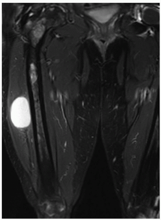
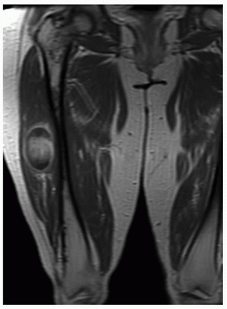
An adult patient presented with long-standing deformity of the right lower extremity. Two coronal MR images, including T2-weighted fat-suppressed and T1-weighted fat-suppressed obtained after intravenous administration of gadolinium, show osseous and soft-tissue abnormalities. Which one of the following is the most likely diagnosis?
 |
 |
A) Osteosarcoma of the proximal femur with soft-tissue metastasis.
B) Mazabraud syndrome.
C) Jaffe-Campanacci syndrome.
D) Rothmund-Thomson syndrome.
E) Werner syndrome.
View Answer
Answer to question 12: B
MR images show a signal abnormality in the right proximal femur extending into the middle of the shaft. In the intertrochanteric region, there are at least two lesions with sclerotic border. In the soft tissue, there is a well-circumscribed lesion showing homogeneous high signal intensity on T2-weighted sequences and rim enhancement on the postcontrast study. All lesions show absence of aggressive features. These findings combined with the clinical history of long-standing deformity are consistent with polyostotic fibrous dysplasia and soft-tissue myxoma, features of Mazabraud syndrome. (A) Osteosarcoma will show more aggressive imaging features, and it extremely rare metastasizes to the soft tissue. Besides, metastatic soft-tissue lesion will not exhibit homogeneous high signal on T2 weighting, but rather heterogeneity with some evidence of bone formation. (C) Jaffe-Campanacci syndrome consists of coexistence of nonossifying fibromas and neurofibromatosis, associated with formation of café-au-lait spots on the skin. (D) Rothmund-Thomson syndrome, also known as congenital poikiloderma, is a hereditary disorder characterized by erythematous and maculopapular skin lesions with areas of hyperpigmentation and coexistence of conventional osteosarcoma. (E) Werner syndrome, also known as adult progeria, is characterized by premature aging, scleroderma-like skin changes, and coexistence of osteosarcomas.
Question 13
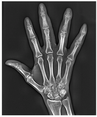
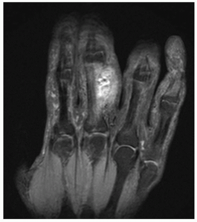
You are shown a radiograph and a coronal T2-weighted MRI of the hand of a 36-year-old woman. Which one of the following is the most likely diagnosis?
 |
 |
A) Ewing sarcoma.
B) Angiosarcoma.
C) Hemangioma.
D) Langerhans cell histiocytosis.
E) Enchondroma.
View Answer
Answer to question 13: C
Radiograph shows a several small radiolucent channels traversing the medullary portion and the cortex of the proximal phalanx of the middle finger associated with a soft-tissue prominence. Coronal T2-weighted fat-suppressed MRI shows high signal intensity lesion affecting both the bone and soft tissue. Low signal intensity foci represent the phleboliths. From the given choices, the most likely diagnosis is an intraosseous hemangioma with soft-tissue extension. (A) Ewing sarcoma is a malignant tumor predominantly affecting the long and flat bones, and involvement of the short tubular bone, although reported in the literature, is extremely rare. (B) The possibility of angiosarcoma should be considered in the differential diagnosis; however, majority of the reported cases were affecting long bones and bones of the pelvis. Involvement of the short tubular bone is unlikely. In addition, the imaging features of angiosarcoma are more aggressive than presented in this case. (D) Langerhans cell histiocytosis is a condition predominantly affecting children, with peak incidence between the ages of 5 and 10 years. The sites of involvement include the axial skeleton and the long bones. Periosteal reaction is usually lamellated. Phleboliths are not the part of presentation. (E) Although the short tubular bones are the common sites of enchondroma, this benign cartilaginous lesion does not extend into the soft tissues, unless it is multifocal such as in Ollier disease.
Question 14
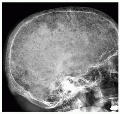
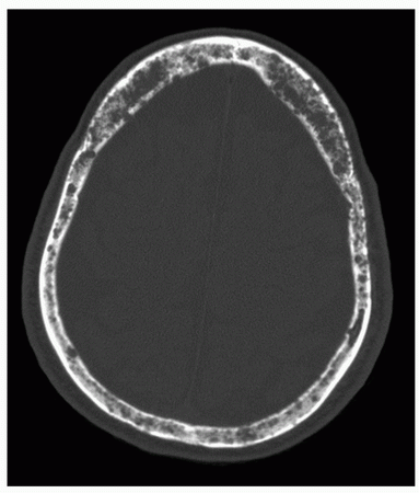
A 76-year-old woman presented with a severe headache. She gives a history of right breast lumpectomy 3 years ago. Based on the lateral radiograph of the skull and axial CT section of the skull, what is your diagnosis?
 |
 |
A) Osteolytic metastases.
B) Hyperparathyroidism.
C) Fibrous dysplasia.
D) Paget disease.
E) Multiple myeloma.
View Answer
Answer to question 14: E
Lateral radiograph of the skull shows numerous variousin-size punched-out lesions, characteristic of multiple myeloma. CT section shows predominant involvement of the diploë, also typical feature for myeloma. (A) Osteolytic metastases to the skull commonly present as a larger areas of destruction. In contrast to myeloma, the inner and outer tables are also affected. (B) Skull abnormalities seen in hyperparathyroidism consist of osteopenia, which gives a characteristic granular appearance of the bones of the vault, referred to as “salt and pepper” appearance, not the punchedout lesions. (C) Fibrous dysplasia affects predominantly the base of the skull in form of areas of sclerosis. (D) Osteolytic phase of Paget disease in the skull is known as osteoporosis circumscripta, appearing as a large area of geographic bone destruction. Mixed and cool phases of the disease affecting the skull manifest as areas of ill-defined focal densities resembling cotton balls.
Question 15
Concerning multiple myeloma, which one of the following statements is NOT true?
A) The skeletal scintigraphy shows invariably increased uptake of the radiopharmaceutical tracer.
B) By immunohistochemistry, the tumor cells show monoclonal expression of lambda light chains.
C) Patients with POEMS syndrome do not have an increased incidence of amyloidosis and rarely have Bence Jones proteinuria.
D) Endosteal scalloping is a characteristic feature of myeloma affecting long tubular bones.
E) A well-differentiated tumors may exhibit almost normalappearing plasma cells.
View Answer
Answer to question 15: A
One of the characteristic features of multiple myeloma, in contrast to metastatic disease, is essentially normal radionuclide bone scan. The remaining statements (B), (C), (D), and (E) are all correct.
Question 16
Concerning polyostotic fibrous dysplasia, which one of the following statements is correct?
A) Histopathology shows woven bone trabeculae surrounded by active osteoblasts.
B) A “shepherd’s crook” deformity of the femur is often the result of multiple pathologic fractures.
C) Skull lesions are seen primarily at the calvaria.
D) Postcontrast MR images show lack of enhancement.
E) McCune-Albright syndrome is caused by gains of chromosomes 1q, 3q, 9q, 11q, and 15q.
View Answer
Answer to question 16: B
One of the characteristic features of polyostotic fibrous dysplasia affecting the femur is so-called shepherd’s crook deformity, which is commonly the result of multiple pathologic fractures. (A) Histopathology of fibrous dysplasia shows immature woven bone trabeculae without osteoblastic rimming, so-called naked trabeculae. (C) Skull lesions in fibrous dysplasia are seen predominantly at the base of the skull in form of increased sclerosis. (D) Postcontrast MRI shows invariably enhancement of the lesions of fibrous dysplasia. (E) Gains of chromosomes 1q, 3q, 9q, 11q, and 15q are characteristic features of multiple myeloma, not polyostotic fibrous dysplasia. McCune-Albright syndrome, which is the association of polyostotic fibrous dysplasia with endocrine disturbances (such as premature sexual development, gigantism or acromegaly, hyperparathyroidism, and Cushing syndrome) and skin pigmentation (cafe-au-lait spots), is caused by somatic mutations in the GNASI gene located in chromosome 20q13.
Question 17
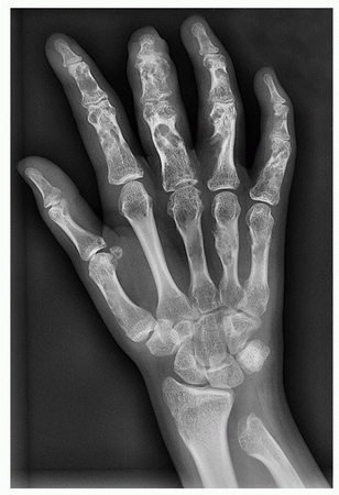
A) Polyostotic fibrous dysplasia.
B) Multiple osteochondromas.
C) Ollier disease.
D) Maffucci syndrome.
E) Neurofibromatosis.
View Answer
Answer to question 17: C
The radiograph of the hand shows multiple sharply marginated radiolucent lesions within the metacarpals and phalanges, some within the cortex, typical for enchondromas. In addition, there is growth stunting of the distal ulna. The combination of these findings is characteristic for Ollier disease. (A) Polyostotic fibrous dysplasia exhibits thinning of the cortex associated with smoky or ground-glass appearance of the medullary portion of bones, without sharply marginated radiolucent lesions typical for enchondromatosis. In addition, overgrowth of the bone rather than growth stunting is occasionally present. (B) The most important identifying features of osteochondromas are that the cortex of the lesion merges without interruption with the cortex of the host bone and that the cancellous portion of the lesion is continuous with the medullary cavity of the adjacent diaphysis, findings not present in the case under consideration. (D) Maffucci syndrome consists of association of Ollier disease with softtissue hemangiomas. The radiography will invariably demonstrate phleboliths, which appear as round calcific foci, not present on the submitted radiograph. (E) Neurofibromatosis may be associated with osseous deformities, characteristic pit-like cortical erosions, and soft-tissue masses, features not present in the case under consideration.
Question 18
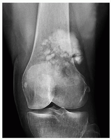
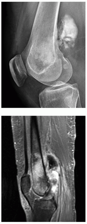
A 19-year-old man presented with pain and mass around the left knee. Based on the radiographs and MRI, what is the most likely diagnosis?
 |
 |
A) Chondrosarcoma.
B) Conventional osteosarcoma.
C) Parosteal osteosarcoma.
D) Periosteal osteosarcoma.
E) Melorheostosis.
View Answer
Answer to question 18: B
Anteroposterior and lateral radiographs show ill-defined sclerotic lesion in the medullary portion of the left distal femur associated with a large, dense soft-tissue mass. Sagittal T1-weighted fat-suppressed MR image obtained after intravenous administration of gadolinium shows significant enhancement of both medullary tumor and soft-tissue mass. Nonenhanced area represents tumor bone formation. The imaging features are typical for conventional osteosarcoma. (A) Chondrosarcoma, although may present as a sclerotic lesion, most of the time exhibits characteristic chondroid calcifications in the form of stippling, flocculent densities, or rings and arcs (“o”s and “c”s). In addition, there is invariably present a deep endosteal scalloping and cortical thickening, absent in the case under consideration. (C) Parosteal osteosarcoma is a surface lesion, usually exhibiting well-defined sclerotic mass outside of the cortex, which is slightly less dense on the periphery. It may possess an incomplete cleft between the tumor and underlying cortex. (D) Periosteal osteosarcoma is also a surface lesion, displaying ill-defined heterogeneous ossifications with calcified spiculations interspersed with areas of radiolucencies representing uncalcified matrix. (E) Melorheostosis, a form of sclerosing dysplasia, may occasionally mimic osteosarcoma, although the surface ossifications, in contrast to osteosarcoma, are well defined, resembling wax dripping on one side of the candle.
Question 19
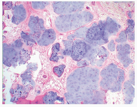
A) Erosion of adjacent bones is a common radiographic finding.
B) Cartilage nodules show small oval or spindled cells.
C) The lesion most commonly arises in the synovium of the joints, bursae, or tendon sheaths.
D) Complete synovectomy is a treatment option.
E) Clinical symptoms include pain, swelling, and stiffness of the joints.
View Answer
Answer to question 19: B
Histopathologic section shows typical appearance of synovial (osteo)chondromatosis. Several cartilaginous nodules are seen covered by a thin layer of synovium. Although the nodules are highly cellular, there is no evidence of spindle cells. All other statements (A), (C), (D), and (E) are correct.
Question 20
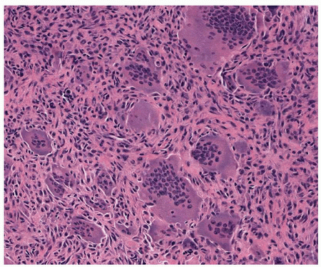
A) The preferential location in the long bone is metaphysis.
B) Most common locations in the skeleton are the distal radius, proximal tibia, distal femur, and proximal humerus.
C) Osteolytic lesion with narrow zone of transition and geographic type of bone destruction are the characteristic radiographic features.
D) It can metastasize to the lung.
E) Most common chromosomal aberration is telomeric associations involving chromosomes 11p, 13p, 14p, 15p, 19q, 20q, and 21p.
View Answer
Answer to question 20: A
Histopathologic section shows the classic features of the giant cell tumor: fibrocytic and monocytic stromal cells and large giant cells containing numerous nuclei. The preferential location of this tumor is the articular end of bone in skeletally mature patient and not in the metaphysis. The remaining statements (B), (C), (D), and (E) are all correct.
Question 21
Which of the following osseous lesion is associated with Jaffe-Campanacci syndrome?
A) Osteochondroma.
B) Osteoid osteoma.
C) Nonossifying fibroma.
D) Osteoblastoma.
E) Fibrous dysplasia.
View Answer
Answer to question 21: C
Jaffe-Campanacci syndrome consists of association of multiple nonossifying fibromas with neurofibromatosis.
Question 22
Which clinical scenario would be most consistent with osteofibrous dysplasia of bone?
A) A 25-year-old woman with swelling of the jaw.
B) A 10-year-old girl with lesion in the distal femur.
C) A 15-year-old boy with lesion in the distal finger.
D) A 65-year-old woman with lesion in the proximal tibia.
E) A 2-year-old boy with cortical lesion in the tibia.
View Answer
Answer to question 22: E
Osteofibrous dysplasia, also known as Kempson-Campanacci lesion, characteristically affects children, with about 60% of the affected patients being younger than 5 years. The preferential site is the anterior cortex of the tibia.
Question 23
Which statement is FALSE regarding mesenchymal chondrosarcoma?
A) Small round tumor cells with low-grade cartilaginous component is characteristic.
B) Cytogenetic studies for t(11;22) are critical to distinguish this tumor from Ewing/PNET sarcoma family.
C) It has indolent behavior with low metastatic rate.
D) Most often involve bones of the jaw and ribs in young adults.
E) Small round tumor cells express positivity for immunohistochemistry stain CD99.
View Answer
Answer to question 23: C
Mesenchymal chondrosarcoma is an aggressive tumor with common distant metastases. The remaining statements (A), (B), (D), and (E) are all correct.
Question 24
Which of the following features are characteristic for clear cell chondrosarcoma?
A) Imaging studies may resemble chondroblastoma.
B) Reactive bone formation may be observed.
C) Large cells with clear or slightly eosinophilic cytoplasm and small nuclei are characteristic.
D) Clear cells of the tumor are positive for S-100 protein.
E) All of the above.
View Answer
Answer to question 24: E
All statements concerning clear cell chondrosarcoma are true.
Question 25
Which of the following lesions histologically resembles an osteoblastoma?
A) Osteochondroma.
B) Osteoid osteoma.
C) Chondroblastoma.
D) Aneurysmal bone cyst.
E) Fracture callus.
View Answer
Answer to question 25: B
Histopathologically, osteoid osteoma and osteoblastoma exhibit similar features. Both lesions show irregular woven bone trabeculae with various degrees of mineralization, osteoblasts and giant cells, ectatic blood vessels, and reactive periosteal shell of new bone. The difference is in size: osteoid osteoma exhibits limited growth potential and size less than 2 cm, whereas osteoblastoma is commonly greater than 2 cm in size.
Question 26
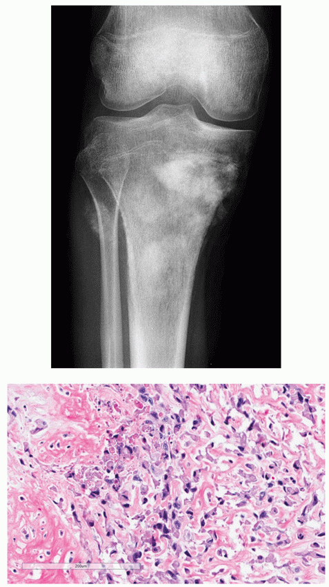
A 13-year-old girl presented with pain in the leg after being kicked during a soccer game. Based on the submitted radiograph and the histopathologic section, what is the most likely diagnosis?
 |
A) Osteoblastoma.
B) Osteoid osteoma.
C) Giant cell-rich osteosarcoma.
D) Conventional osteosarcoma.
E) Healing fracture of the proximal tibia.
View Answer
Answer to question 26: D
Anteroposterior radiograph of the right knee shows fluffy sclerotic densities in the metaphysis and proximal diaphysis of the tibia representing tumor bone formation, associated with velvet-type periosteal reaction. Similar densities are seen within the soft-tissue mass at the site of the proximal fibula. These are the typical imaging features of conventional osteosarcoma. The diagnosis is confirmed by the histopathologic section, showing markedly pleomorphic osteoblastic tumor cells with hyperchromatic nuclei forming lace-like osteoid. (A) Imaging features of osteoblastoma occasionally may mimic those of osteosarcoma; however, the lesion exhibits more focally confined densities, and the periosteal reaction is solid, not interrupted as in the case under consideration. Histopathology shows large but benign osteoblasts forming immature woven bone trabeculae. (B) Osteoid osteoma shows resemblance to osteoblastoma but exhibits completely different imaging features than osteosarcoma. The size of the lesion is usually less than 2 cm, and periosteal reaction is of solid type. Moreover, the lesion does not produce the soft-tissue mass. Histopathology shows a well-defined nidus composed of osteoid and trabeculae of immature woven bone, surrounded by sclerotic reactive bone. (C) Imaging features of giant cell-rich osteosarcoma may be indistinguishable from that of conventional osteosarcoma; however, histopathology shows striking resemblance to giant cell tumor with numerous osteoclast-like giant cells and scant osteoid formation, unlike the histopathologic section under consideration. (E) Callus formation of healing fracture shows more organized pattern on imaging studies, and there is no softtissue mass present. Furthermore, histopathology shows lack of malignant cells and lack of tumor bone or tumor osteoid.
Question 27
Concerning lipoma arborescens, which of the following statements is (are) true?
A) The lesion is more prevalent in males, usually in the fourth and fifth decades.
B) Imaging features are not characteristic, and only biopsy is diagnostic.
C) MRI shows high signal intensity on T1-weighted sequences.
D) A and C.
E) All of the above.
View Answer
Answer to question 27: D
Lipoma arborescent is a rare intra-articular disorder characterized by a nonneoplastic lipomatous proliferation of the synovium resembling a frond-like mass. It is more prevalent in males and usually diagnosed in the fourth and fifth decades of life. MRI features are characteristic and diagnostic for this disorder, showing fat signal intensity on all imaging sequences.
Question 28
Concerning synovial (osteo)chondromatosis, which of the following statements is (are) true?
A) The knee is a preferential site.
B) Joint effusion is a common clinical finding.
C) Histopathology shows pleomorphic cartilaginous cells arranged in clusters, exhibiting dark nuclei.
D) A and C.
E) All of the above.
View Answer
Answer to question 28: E
Synovial (osteo)chondromatosis is marked by the metaplastic proliferation of multiple cartilaginous nodules in the synovial membrane of the joints, bursae, or tendon sheaths. The knee joint is a preferential site of involvement. Joint effusion, tenderness, limited motion in the joint, and a softtissue mass are common clinical findings. Histopathology shows numerous highly cellular cartilaginous nodules. The cells with plump and dark nuclei are arranged in clusters and show moderate pleomorphism.
Question 29
A 65-year-old man presented with pain in the thigh. Based on the submitted radiograph, CT, and histopathologic section, what is the most likely diagnosis?
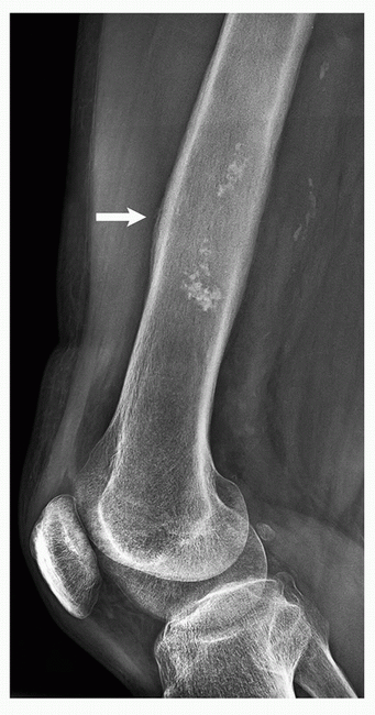 |
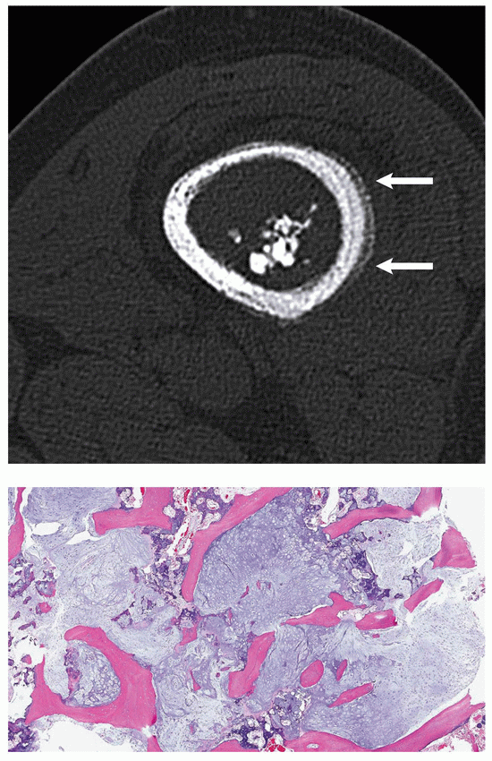 |
A) Enchondroma.
B) Conventional chondrosarcoma.
C) Medullary bone infarct.
D) Chondromyxoid fibroma.
E) Chondroblastoma.
View Answer
Answer to question 29: B
Lateral radiograph of the distal femur shows intramedullary lesion with wide zone of transition and chondroid calcifications. In addition, noted is focal cortical thickening and periosteal reaction (arrows). These findings are also demonstrated on the axial CT section through the tumor. The histopathology shows pleomorphic tumor with hyperchromatic nuclei permeating the bone trabeculae. The imaging findings and histopathology are characteristic for conventional intramedullary chondrosarcoma. (A) Although enchondroma also may display chondroid calcifications, generally there is no periosteal reaction, unless the pathologic fracture has occurred. Also, the cortex is not thickened; to the contrary, most of the time noted is thinning of the cortex. (C) Medullary bone infarction also exhibits calcifications, but in contrast to the chondroid tumors, the calcifications are coarser, and peripheral serpentine sclerotic fibro-osseous wall is commonly observed. Moreover, the cortex is not thickened. (D) Chondromyxoid fibroma exhibits narrow zone of transition and lack of visible calcifications. (E) Chondroblastoma is a tumor of immature skeleton, invariably located within an epiphysis. It exhibits narrow zone of transition and lack of periosteal reaction at the site of the lesion.
Question 30
Which one of the following statements concerning chondromyxoid fibroma is NOT true?
A) The radiography shows typical chondroid calcifications.
B) Narrow zone of transition and geographic type of bone destruction are the characteristic imaging features.
C) Tibia is a preferential site of the lesion.
D) Histopathology shows lobulated areas of spindle-shaped or stellate cells distributed within abundant myxoid stroma.
E) Imaging appearance may be similar to an aneurysmal bone cyst.
View Answer
Answer to question 30: A
Chondromyxoid fibroma is the only cartilaginous lesion that does not show visible calcifications on radiography. The remaining statements (B), (C), (D), and (E) are all correct.
Question 31
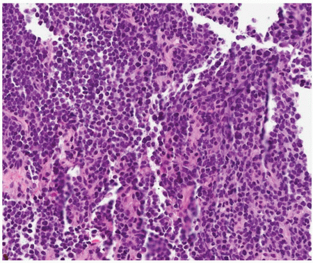
A) Langerhans cell histiocytosis.
B) Ewing sarcoma.
C) Large B-cell lymphoma.
D) Mesenchymal chondrosarcoma.
E) Multiple myeloma.
View Answer
Answer to question 31: B
The histopathologic section shows uniformly arranged small round cells exhibiting indistinguished borders, with darkly stained round nuclei, and scanty eosinophilic cytoplasm, highly characteristic for Ewing sarcoma. (A) Langerhans cell histiocytosis generally shows large pale histiocytes (Langerhans cells) and giant cells, as well as numerous eosinophilic granulocytes. The nuclei exhibit characteristic longitudinal grooves. (C) Large B-cell lymphoma exhibits pleomorphic round cells with well-defined cytoplasmic outline and enlarged, occasionally cleaved nuclei with nucleoli. (D) Mesenchymal chondrosarcoma on histopathologic examination shows two components: small, round uniform-sized cells of mesenchymal tissue with round or ovoid nuclei, which resemble Ewing sarcoma, and chondroblastic tissue with small spindle cells. (E) Multiple myeloma shows plasma cells with eccentric nuclei and basophilic cytoplasm.
Question 32
Concerning multiple myeloma, which of the following statements is (are) true?
A) Most of the time the scintigraphy is normal.
B) Sclerosing variant is the part of POEMS syndrome.
C) Endosteal scalloping is a common radiographic feature.
D) By immunohistochemistry, the tumor cells show a monoclonal expression of lambda light chains.
E) All statements are true.
View Answer
Answer to question 32: E
All statements concerning multiple myeloma are true.
Question 33
Which of the following statements is inconsistent with diagnosis of periosteal osteosarcoma?
A) Cartilaginous matrix is a major component of the tumor.
B) It is more aggressive than parosteal osteosarcoma.
C) Belongs to the group of surface tumors.
D) The majority of the tumors affect the tibia and the femur.
E) MRI shows characteristic fluid-fluid levels.
View Answer
Answer to question 33: E
Fluid-fluid levels seen on MRI are characteristic features of telangiectatic osteosarcoma and not periosteal osteosarcoma. All remaining statements are correct.
Question 34
Which of the following lesions does not contain giant cells?
A) Giant cell tumor.
B) Nonossifying fibroma.
C) Hemangioma.
D) Aneurysmal bone cyst.
E) Brown tumor of hyperparathyroidism.
View Answer
Answer to question 34: C
All listed lesions except of hemangioma contain giant cells.
Question 35
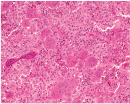
A) Lamellated periosteal reaction is a common finding.
B) Imaging features may resemble osteomyelitis.
C) The neoplastic cells are positive for CD45 and CD68.
D) “Floating teeth” is a characteristic feature.
E) Electron microscopy shows characteristic Birbeck granules.
View Answer
Answer to question 35: C
The histopathologic section shows densely arranged large pale histiocytes (Langerhans cells) as well as giant cells and eosinophilic granulocytes, characteristic of Langerhans cell histiocytosis. The tumor cells are positive for S-100 protein and CD1a but not for CD45 and CD68. The remaining statements concerning Langerhans cell histiocytosis are all correct.
Question 36
Which one of the following lesions generally does not produce periosteal reaction?
A) Chondromyxoid fibroma.
B) Aneurysmal bone cyst.
C) Langerhans cell histiocytosis.
D) Osteoblastoma.
E) Simple bone cyst.
View Answer
Answer to question 36: E
All listed lesions except of a simple bone cyst are associated with a periosteal reaction.
Question 37
Which one of the following lesions generally does not exhibit matrix mineralization?
A) Osteoid osteoma.
B) Giant cell tumor.
C) Chondroblastoma.
D) Enchondroma.
E) Clear cell chondrosarcoma.
View Answer
Answer to question 37: B
Giant cell tumor invariably does not exhibit any matrix mineralization. In contrast, osteoid osteoma may show mineralization of the nidus, and chondroblastoma, enchondroma, and clear cell chondrosarcoma all show chondroid calcifications.
Question 38
Concerning desmoplastic fibroma, which of the following statements is FALSE?
A) It shows always a narrow zone of transition.
B) It is a locally aggressive lesion.
C) Histopathology shows densely collagenized hypocellular stroma containing spindle-shaped fibroblasts.
D) Honeycomb pattern of bone destruction is commonly observed.
E) Histopathologically, the lesion may resemble periosteal desmoid.
View Answer
Answer to question 38: A
Desmoplastic fibroma is a locally aggressive lesion characterized by formation of abundant collagen fibers by the tumor cells. Radiographically, it exhibits the expansive features, usually with nonsclerotic but sharply defined borders, although occasionally a wide zone of transition is present. The remaining statements are all correct.
Question 39
Which one of the following lesions may be associated with a soft-tissue mass?
A) Aneurysmal bone cyst.
B) Simple bone cyst.
C) Giant cell tumor.
D) A and C only.
E) All of the above.
View Answer
Answer to question 39: D
Aneurysmal bone cyst and giant cell tumor, although both the benign lesions, may be occasionally associated with a soft-tissue mass. To the contrary, a simple bone cyst never exhibits a soft-tissue extension.
Question 40
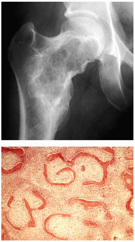
A) Osteofibrous dysplasia.
B) Fibrous dysplasia.
C) Fibrosarcoma.
D) Desmoplastic fibroma.
E) Giant cell tumor.
View Answer
Answer to question 40: B
The anteroposterior radiograph of the right hip shows a radiolucent lesion with narrow zone of transition and sclerotic border (rind sign) within the femoral neck. There is no evidence of a periosteal reaction. The histopathologic section shows irregularly shaped immature woven bone trabeculae resembling Chinese characters, with conspicuous absence of osteoblastic rimming (so-called naked trabeculae), diagnostic of fibrous dysplasia. (A) Osteofibrous dysplasia affects predominantly the tibia, and histopathology, in contrast to fibrous dysplasia, shows prominent osteoblastic rimming of the bone trabeculae (“dressed” trabeculae). (C) Fibrosarcoma is an aggressive tumor showing permeative or moth-eaten type of bone destruction and periosteal reaction. Histopathology shows pleomorphic spindle cells arranged in long fascicles forming characteristic “herringbone tweed” pattern. (D) Desmoplastic fibroma, although may exhibit a narrow zone of transition, usually have a nonsclerotic border. The histopathology shows densely collagenized hypocellular stroma that contains spindle-shaped fibroblasts. (E) Giant cell tumor invariably extends into the articular end of bone and shows lack of sclerotic border. Histopathology shows characteristic dual population of stromal cells (fibrocytic or monocytic) and of giant cells uniformly distributed throughout the lesion.
Question 41
Concerning osteofibrous dysplasia, which one of the following statements is true?
A) Cytogenetic studies revealed trisomies for chromosomes 7, 8, 12, and 22.
B) Bone trabeculae show appositional activity by polarized osteoblasts.
C) Anterior aspect of the tibia is a preferential location.
D) It may exhibit similar imaging and histopathologic features to the adamantinoma of the long bones.
E) All statements are correct.
View Answer
Answer to question 41: E
All statements concerning osteofibrous dysplasia are correct.
Question 42
Regarding polyostotic fibrous dysplasia, which one of the following statements is NOT true?
A) It may be associated with hyperparathyroidism.
B) Massive formation of cartilage may be observed.
C) Osteoblastic activity is a characteristic feature.
D) Skull lesions affect primarily the base.
E) It may be part of Mazabraud syndrome.
View Answer
Answer to question 42: C
In contrast to osteofibrous dysplasia, the characteristic histopathologic feature of fibrous dysplasia is the presence of immature woven bone trabeculae without osteoblastic activity (“naked” trabeculae). The remaining statements are all true.
Question 43
Concerning synovial sarcoma, which one of the following statements is FALSE?
A) It arises from the synovial membrane of the joint.
B) A translocation involving chromosomes × and 18 [t(X;18)(p11;q11) is a common feature.
C) MRI shows fluid-fluid levels in 18% of patients.
D) It shows positivity for CD99 and cytokeratins.
E) The tumor occurs usually before the age of 50.
View Answer
Answer to question 43: A
Synovial sarcoma is an uncommon malignant mesenchymal neoplasm. Despite its name, it does not arise from synovium. It occurs mainly in para-articular regions close to joint capsule, bursae, and tendon sheets. The remaining statements are all true.
Question 44
Regarding fibrosarcoma, which of the following statements is true?
A) Radiography shows a sclerotic lesion associated with periosteal reaction.
B) Histopathology shows characteristic herringbone tweed pattern of arrangement of the malignant cells.
C) The spindle cells have normochromatic nuclei with decreased ratio of nucleus to cytoplasm.
D) Fluid-fluid levels on MRI are commonly encountered.
E) It is a common lesion in the pediatric age.
View Answer
Answer to question 44: B
The characteristic histopathologic feature of fibrosarcoma is “herringbone tweed” pattern of arrangement of the malignant pleomorphic spindle cells. (A) Radiography shows an osteolytic lesion with wide zone of transition and no evidence of sclerosis. (C) The spindle cells show hyperchromatic nuclei with increased ratio of nucleus to cytoplasm. (D) Fluid-fluid levels on MRI are not the feature of fibrosarcoma. (E) Fibrosarcoma usually occurs in the third to sixth decade of life, and only a few cases have been reported in children.
Question 45
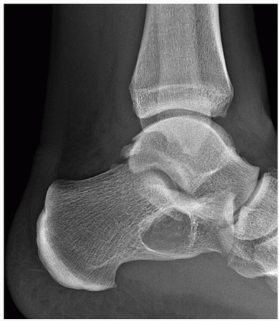
A 45-year-old man injured his ankle. Based on this lateral radiograph, what is the most likely diagnosis?
 |
A) Posttraumatic cyst.
B) Intraosseous lipoma.
C) Intraosseous ganglion.
D) Simple bone cyst.
E) Aneurysmal bone cyst.
View Answer
Answer to question 45: D
The lateral radiograph of the ankle shows a radiolucent lesion with narrow zone of transition and geographic type of bone destruction, located within the Ward triangle of the calcaneus. It has all characteristic features of a simple bone cyst and typical location in this portion of calcaneus. (A) Posttraumatic cyst is uncommon lesion, and usually appears as the result of a fracture about 2 months after injury. These lesions were reported in children, the radius being the usual site. (B) Intraosseous lipoma has a very similar imaging feature as a simple bone cyst; however, it shows invariably a central mineralization (ossifications or calcifications). (C) Intraosseous ganglion is commonly located in a long tubular bone, close to the joint. (E) The calcaneus is an unusual site of aneurysmal bone cyst. Moreover, this lesion commonly shows expansive features and periosteal reaction.
Question 46
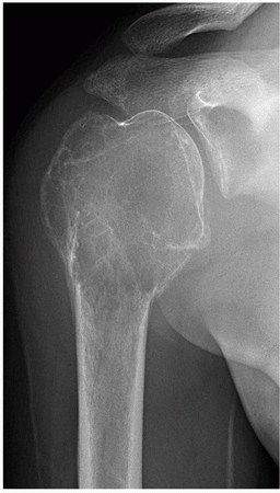
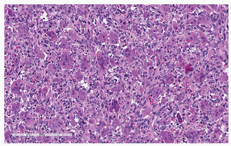
A 35-year-old woman presented with right shoulder pain of 4 months’ duration. Based on the submitted radiograph and histopathologic section, what is the most likely diagnosis?
 |
 |
A) Simple bone cyst.
B) Aneurysmal bone cyst.
C) Giant cell tumor.
D) Plasmacytoma.
E) Metastasis from renal cell carcinoma.
View Answer
Answer to question 46: C
The radiograph of the right shoulder shows a radiolucent lesion affecting proximal humerus and extending into the articular end of the bone. There is evidence of a prior pathologic fracture at the proximal part with associated well-organized periosteal reaction. No obvious soft-tissue mass is present. There is absence of mineralization. The histopathologic section shows numerous osteoclast-like giant cells uniformly scattered among round and spindle-shaped mononuclear tumor cells. The most likely diagnosis in the patient of this age is a giant cell tumor. (A) Although a proximal humerus is a common site of the simple bone cyst, these lesions invariably are metaphyseal or diaphyseal in location and do not extend into the subchondral part of bone, as giant cell tumor does. Moreover, majority of these lesions occur in children. (B) Imaging features of aneurysmal bone cyst may mimic those of a giant cell tumor; however, similarly to the simple bone cyst, these lesions occur predominantly in children and young adults. Furthermore, solid buttress of periosteal reaction is a common feature in the long tubular bones. (D) Plasmacytoma usually presents as a radiolucent lesion and at times may affect the articular end of a long bone. The tumor, however, usually is seen in much older group of patients, commonly between the fifth and seventh decades. (E) Lytic metastases from renal cell carcinoma may mimic the appearance of a giant cell tumor; however, involvement of the articular end of bone is unusual.
Question 47
Regarding simple bone cyst, which one of the following statements is FALSE?
A) A “fallen fragment” sign is a characteristic radiographic feature.
B) Most of the lesions occur in the proximal humerus and proximal femur.
C) About 65% of the cysts occur in teenagers.
D) In the calcaneus, it is located within the Ward triangle.
E) Periosteal reaction is a common feature.
View Answer
Answer to question 47: E
Simple bone cyst typically shows no periosteal reaction, unless a pathologic fracture has occurred. The remaining statements are all true.
Question 48
One of the following lesions DOES NOT affect the spine:
A) Osteoid osteoma.
B) Osteoblastoma.
C) Hemangioma.
D) Osteofibrous dysplasia.
E) Aneurysmal bone cyst.
View Answer
Answer to question 48: D
All of the listed conditions may affect the spine, except for osteofibrous dysplasia, which commonly affects the tibia.
Question 49
Concerning Hodgkin lymphoma, all of the following statements are true EXCEPT one:
A) “Ivory vertebra” is a common finding.
B) Reed-Sternberg cells are positive for CD30.
C) Postcontrast MRI shows enhancement of the lesion.
D) Skeletal scintigraphy shows absence of uptake of the radiopharmaceutical tracer.
E) Fibrous stroma with mixed cellular infiltrate of small round cells and large histiocytes is characteristic for this lesion.
View Answer
Answer to question 49: D
Hodgkin lymphoma invariably shows increased uptake of the radiopharmaceutical tracer. The remaining statements are all correct.
Question 50
All of the following conditions should be included in the differential diagnosis of osteoid osteoma, EXCEPT for which one?
A) Stress fracture.
B) Osteoblastoma.
C) Brodie abscess.
D) Enostosis.
E) Parosteal osteosarcoma.
View Answer
Answer to question 50: E
Parosteal osteosarcoma has characteristic imaging features, most notably a homogenously sclerotic ivory mass attached to the cortex of a long or flat bone. Conversely, all the other listed conditions may occasionally mimic the features of osteoid osteoma. (A) Stress fracture may be mistaken for cortical osteoid osteoma, although linear radiolucency runs perpendicular or at angle to the cortex, whereas the radiolucent nidus of osteoid osteoma is round or oblong. (B) Osteoblastoma may look exactly like osteoid osteoma, except for its size: while the nidus of osteoid osteoma measures usually 1.5 cm or less, the lesion of osteoblastoma is commonly 2.0 cm or larger. (C) Brodie abscess may mimic medullary osteoid osteoma, the important differential feature being the commonly present serpentine radiolucent tract extending from the abscess. (D) Enostosis, also known as a bone island, may mimic medullary osteoid osteoma. The important diagnostic feature of the former lesion includes distinctive radiating streaks (thorny radiation, pseudopodia) forming a rough border.
Question 51
Which one of the following lesions histopathologically resembles an osteoblastoma?
A) Osteochondroma.
B) Osteoid osteoma.
C) Chondroblastoma.
D) Aneurysmal bone cyst.
E) Fracture callus.
View Answer
Answer to question 51: B
Histopathologically, osteoid osteoma and osteoblastoma show very similar features. Both lesions show irregular woven bone with various degrees of mineralization, osteoblasts and giant cells, ectatic blood vessels, and reactive periosteal shell of new bone. The difference is in size: osteoid osteoma has a limited growth, and usual size is 1.0 to 1.5 cm, whereas osteoblastoma is greater than 2.0 cm in size.
Question 52
Which one of these patients most likely has a metastatic lesion?
A) A 32-year-old woman with permeative lesion in the proximal femur exhibiting an interrupted periosteal reaction.
B) A 72-year-old man with osteolytic lesion in the proximal right humerus exhibiting a narrow zone of transition.
C) A 75-year-old woman with osteolytic lesion in the proximal left humerus exhibiting a wide zone of transition, an interrupted periosteal reaction, and a large soft-tissue mass.
D) A 68-year-old man with punched-out lesions in the skull.
E) An 8-year-old girl with osteolytic lesion in the left tibia, exhibiting wide zone of transition and lamellated type of periosteal reaction.
View Answer
Answer to question 52: B
In this age, any osseous lesion, even the one having a “benign” appearance, should be considered a metastasis, unless proven otherwise.
(A) This patient is not in the “metastatic age group,” and, in addition, the metastatic lesions usually do not exhibit periosteal reaction. With the features described, the patient most likely has a primary malignant bone tumor. (C) Although this patient is in the “metastatic age group,” the presence of periosteal reaction and a large soft-tissue mass point to the diagnosis of a primary malignant bone tumor. (D) Punched-out lesions in the skull are classic features of multiple myeloma. (E) In this age, the osteolytic lesion with wide zone of transition and lamellated periosteal reaction may represent Ewing sarcoma, Langerhans cell histiocytosis, or even osteomyelitis but not a metastasis.
Question 53
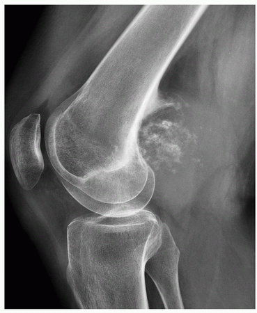
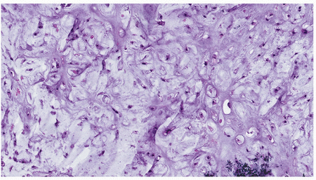
A 39-year-old man presented with a large mass in the popliteal region of the knee of several months’ duration. He experienced only mild discomfort but noticed a slow increase in the size of the mass. Based on the submitted radiograph and histopathologic section, what is the most likely diagnosis?
 |
 |
A) Parosteal osteosarcoma.
B) Periosteal chondroma.
C) Periosteal chondrosarcoma.
D) Conventional chondrosarcoma.
E) Tumoral calcinosis.
View Answer
Answer to question 53: C
The lateral radiograph of the knee shows a large soft-tissue mass with chondroid calcifications eroding but not invading the posterior cortex of the distal femur and evoking a periosteal reaction. These are the imaging features of periosteal chondrosarcoma. The diagnosis is confirmed by histopathologic section showing hyaline cartilage with pleomorphic tumor cells and hyperchromatism of the nuclei consistent with intermediate-grade malignancy. (A) The posterior aspect of the distal femur is a common site of parosteal osteosarcoma; however, this tumor presents on radiography as a homogenously sclerotic mass attached to the cortex. (B) Periosteal chondroma may exhibit similar radiographic features as periosteal chondrosarcoma; the lesion, however, is much smaller, and the distal femur is not a preferential site, the most common location being the proximal humerus. (D) Conventional chondrosarcoma is a medullary lesion, although it may extend into the soft tissues. The tumor under consideration is a surface lesion, without intramedullary involvement. (E) Tumoral calcinosis is characterized by the presence of single or multiple periarticular lobulated cystic masses containing chalky material. The masses are painless, usually occur in children and adolescents, and most commonly are formed around the shoulders, hips, and elbow joints, and on the extensile surfaces of the limbs.
Question 54
Which one of the following features is characteristic for clear cell chondrosarcoma?
A) Commonly epiphyseal lesion.
B) Clear cytoplasm.
C) Woven bone.
D) Clear cells are positive for S-100 protein.
E) All of the above.
View Answer
Answer to question 54: E
Concerning clear cell chondrosarcoma, all of the statements are true.
Question 55
What is the characteristic neoplastic cell of a chordoma?
A) Chondrocyte.
B) Myocyte.
C) Neuroglia.
D) Osteoblast.
E) Physaliphorous cell.
View Answer
Answer to question 55: E
The chordoma consists of cord-like arrays and lobules of large polyhedral cells with a vacuolated cytoplasm and vesicular nuclei, referred to as physaliphorous cells (from Greek for “bubble bearing”).
Question 56
An 82-year-old man presented with lower back pain. Radiographic examination demonstrated “missing” pedicle of L4 vertebra, without additional abnormalities. What is the most likely diagnosis?
A) Metastasis.
B) Congenital absence of the pedicle.
C) Aneurysmal bone cyst.
D) Osteoblastoma.
E) Plasmacytoma.
View Answer
Answer to question 56: A
In the older patient presenting with back pain, the absence of the pedicle (the “missing pedicle” sign) owing to bone destruction should always be regarded as a metastasis, unless proven otherwise. (B) Congenital absence of the pedicle is usually diagnosed in much younger age, and the tip-off is the hypertrophy of the contralateral pedicle. The condition is usually asymptomatic. (C) Posterior elements of the vertebra are the common site of an aneurysmal bone cyst; the condition, however, occurs in much younger age group, most of the cases diagnosed between ages 5 and 20. In addition, aneurysmal bone cyst usually exhibits expansive imaging features, not present in the case under consideration. (D) Whenever a radiolucent lesion is present in the posterior elements of the vertebra, osteoblastoma should be considered in the differential diagnosis; however, similar to the aneurysmal bone cyst, it occurs in much younger age group, majority of the patients being between 10 and 35 years of age. In addition, fluffy densities are usually present within the radiolucent focus. (E) The vertebra is the usual site of plasmacytoma; however, unlike the metastatic lesion, the vertebral body not the pedicle is the site of destruction.
Question 57
What description is most consistent with a malignant bone tumor?
A) Radiolucent lesion with geographic type of bone destruction and a narrow zone of transition.
B) Radiolucent lesion with moth-eaten bone destruction, without periosteal reaction.
C) Radiolucent lesion with geographic type of bone destruction and a soft-tissue mass.
D) Radiolucent lesion with geographic type of bone destruction, narrow zone of transition, and a solid buttress of periosteal reaction.
E) Sclerotic lesion with a rough border, exhibiting thorny radiations and a positive radionuclide bone scan.
View Answer
Answer to question 57: B
From the given choices, the radiolucent lesion exhibiting moth-eaten type of bone destruction, despite lack of a periosteal reaction, most likely represents a malignant bone tumor. (A) Geographic type of bone destruction and a narrow zone of transition are the hallmarks of a nonaggressive, most likely benign bone lesion. (C) Soft-tissue mass may be associated with benign bone lesion, such as an aneurysmal bone cyst, giant cell tumor, or desmoplastic fibroma. It is the geographic type of bone destruction that usually (although not always) indicates benignity of the lesion. (D) Again, geographic type of bone destruction, narrow zone of transition, and uninterrupted, solid buttress of periosteal reaction are associated with benign lesions, such as an aneurysmal bone cyst or chondromyxoid fibroma. (E) Sclerotic lesion with a rough border, exhibiting thorny radiations, is most likely a benign bone island (enostosis). These lesions occasionally demonstrate on skeletal scintigraphy an increased uptake of radiopharmaceutical tracer secondary to increased metabolic and osteoblastic activity.
Question 58
Which one of the following markers is most specific for the diagnosis of chordoma?
A) S-100 protein.
B) Cytokeratins.
C) Brachyury.
D) CEA.
E) EMA.
View Answer
Answer to question 58: C
Although chordoma is positive for (A) S-100 protein, (B) cytokeratins, (D) carcinoembryonic antigen (CEA), and (E) epithelial membrane antigen (EMA), brachyury, a protein encoded by T gene, has been shown to be the most specific marker for this tumor, rendering specificity above 90%.
Question 59
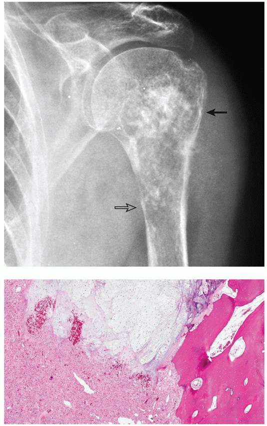
A 72-year-old woman presented with pain in the upper left arm. Based on the submitted radiograph and histopathologic section, what is the most likely diagnosis?
 |
A) Mesenchymal chondrosarcoma.
B) Dedifferentiated chondrosarcoma.
C) Undifferentiated pleomorphic sarcoma.
D) Enchondromatosis.
E) Chondroblastic osteosarcoma.
View Answer
Answer to question 59: B
The radiograph shows a mixed radiolucent and sclerotic lesion in the proximal humerus. The proximal part of the lesion shows chondroid calcifications and intact cortex (arrow), suggesting a slow-growing tumor. The distal part of the lesion demonstrated more aggressive appearance, osteolytic destruction of medullary portion of bone, and cortical invasion (open arrow). These are the imaging features of dedifferentiated chondrosarcoma, confirmed on the histopathologic examination. Observe biphasic character of the tumor with low-grade cartilaginous component (top) juxtaposed to the noncartilaginous high-grade sarcoma (bottom and left), invading the bone trabeculae (right). High-grade sarcoma may consist of osteosarcoma, malignant fibrous histiocytoma, fibrosarcoma, rhabdomyosarcoma, or undifferentiated sarcoma. (A) Mesenchymal chondrosarcoma histopathologically also exhibits biphasic pattern, which consists of two components: mesenchymal tissue made up of small, round uniform-sized cells with round or ovoid nuclei and well-differentiated cartilaginous matrix containing foci of calcifications and occasionally metaplastic bone. (C) Undifferentiated pleomorphic sarcoma is not a biphasic tumor. It is characterized by marked cytologic and nuclear pleomorphism, often with bizarre-appearing giant cells admixed with spindle and histiocyte-like cells. A storiform growth pattern and stromal chronic inflammatory cells are commonly present. (D) Enchondromatosis demonstrates completely different imaging and histopathologic features. Although as a component of Ollier disease, the condition may show some aggressive appearance, generally the radiolucent masses of cartilage with foci of calcifications closely resemble solitary enchondromas. Histopathologically, the lesions in enchondromatosis are essentially indistinguishable from those of a solitary enchondromas, although they sometimes tend to be myxoid, more cellular, and may exhibit more cellular atypia. (E) Chondroblastic osteosarcoma usually displays a fairly well-differentiated cartilaginous matrix, often with only moderate cytologic signs of malignancy but without the typical lobular pattern of the hyaline cartilage seen in well-differentiated chondrosarcoma. A characteristic feature of this tumor is the indistinct transition from the cartilaginous matrix to osseous matrix, unlike in dedifferentiated chondrosarcoma, where the transition is abrupt.
Question 60
Which one of the following statements concerning chordoma is NOT true?
A) Cord-like arrays and lobules of large polyhedral cells with a vacuolated “bubbly” cytoplasm is a characteristic feature.
B) Loss of chromosomes 3, 4, 10, and 13 has been reported.
C) Partial or complete PTEN gene deficiency.
D) Clivus is one of the preferential sites.
E) Occurs only at sites in which notochordal remnants are normally present.
View Answer
Answer to question 60: E
Chordoma occurs only at sites in which notochordal remnants are NOT normally present. Therefore, although it is a midline lesion of the axial skeleton, it never occurs in the nuclei pulposi. The statements A, B, C, and D are all correct.
Question 61
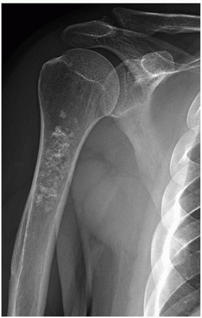
A 42-year-old woman injured her right shoulder in a skiing accident. Based on the submitted radiograph, what is the most likely diagnosis?
 |
A) Medullary bone infarct.
B) Conventional chondrosarcoma.
C) Clear cell chondrosarcoma.
D) Chondromyxoid fibroma.
E) Enchondroma.
View Answer
Answer to question 61: E
The radiograph shows a radiolucent lesion within the medullary portion of the proximal humerus containing variousin-size chondroid calcifications. Observe that the cortex at the site of the lesion is not thickened, there is no evidence of endosteal scalloping and no periosteal reaction. Thickened cortex distal to the lesion represents deltoid tuberosity. The described findings are characteristic features of enchondroma. (A) Medullary bone infarct also exhibits calcifications and therefore may resemble an enchondroma. The calcifications, however, are coarser and demarcated from the viable bone by serpentine fibro-osseous membrane. (B) Imaging features of low-grade chondrosarcoma occasionally can mimic those of enchondroma; however, there is focal thickening of the cortex and the presence of deep endosteal scalloping. (C) Clear cell chondrosarcoma preferentially affects the articular ends (epiphysis) of long tubular bones. On radiography, the lesion is predominantly lytic, sometimes expansive, commonly with a sclerotic border, and occasionally with central calcifications or ossifications. Endosteal scalloping may be present. The imaging features resemble chondroblastoma. (D) Chondromyxoid fibroma is an eccentric lesion, exhibiting narrow zone of transition and geographic type of bone destruction. It commonly is associated with a buttress of solid periosteal reaction. Unlike most of enchondromas, it does not display visible chondroid calcifications.
Question 62
Concerning chondromyxoid fibroma, all the following statements are correct EXCEPT which one?
A) Chondroid calcifications are a common feature.
B) Eccentric location within the bone.
C) It may resemble an aneurysmal bone cyst.
D) Periosteal reaction is a common finding in a long bone.
E) Giant cells are a part of histopathologic presentation.
View Answer
Answer to question 62: A
Chondromyxoid fibroma DOES NOT exhibit visible chondroid calcifications. The remaining statements are all true.
Question 63
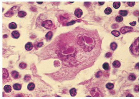
A 45-year-old man was admitted to the hospital with severe back pain. Radiography demonstrated a sclerotic L2 vertebra. An open biopsy was performed. Based on the submitted histopathologic section, what is the most likely diagnosis?
 |
A) Plasmacytoma.
B) Hodgkin lymphoma.
C) Large B-cell lymphoma.
D) Langerhans cell histiocytosis.
E) Metastasis from thyroid carcinoma.
View Answer
Answer to question 63: B
The histopathologic section shows in addition to pleomorphic round cells, the characteristic for Hodgkin lymphoma binucleate Reed-Sternberg cell with prominent eosinophilic nucleoli. Sclerotic vertebrae are common findings in Hodgkin lymphoma. (A) Plasmacytoma exhibits almost invariably osteolytic and not sclerotic features. Moreover, the histopathology shows sheets of atypical plasmacytoid cells. The round nuclei possess a dense, coarse chromatin that has a typical cartwheel-like distribution. (C) Large B-cell lymphoma exhibits pleomorphic round cells with well-defined cytoplasmic outline and enlarged cleaved nuclei with prominent nucleoli. The cytoplasm is basophilic. (D) Langerhans cell histiocytosis may affect the vertebrae. The characteristic finding is so-called vertebra plana, which represents collapsed of vertebral body secondary to destruction of bone by granulomatous lesion. Histopathology shows agglomerates of more or less densely arranged large rounded cells without cytoplasmic extensions. The characteristic Langerhans cells can be specifically identified in electron microscopy by the presence of racquet-shaped cytoplasmic organelles known as Birbeck granules. (E) Metastases from thyroid carcinoma are invariably lytic, not sclerotic. Histopathology may show foci of papillary formation and follicular elements with cells containing clear nuclei with pseudoinclusions.
Question 64
All of the following tumors may metastasize to the lungs, EXCEPT for which one?
A) Telangiectatic osteosarcoma.
B) Giant cell tumor.
C) Parosteal osteosarcoma.
D) Chondroblastoma.
E) Synovial sarcoma.
View Answer
Answer to question 64: C
Parosteal osteosarcoma is a slow-growing, low-grade tumor that affects only surface of the bone and therefore has the much better prognosis than do the other types of osteosarcomas. It does not produce distant metastases. To the contrary, the other listed lesions, some even benign, may metastasize to the lungs. (A) Telangiectatic osteosarcoma, one of the most aggressive types of osteosarcomas, commonly produces lung metastases. (B) Giant cell tumor, even a benign variant, also may metastasize to the lungs. According to various reports, about 2% of the patients with this lesion develop pulmonary metastases on average 3 to 4 years after diagnosis. It is of interest, that these metastatic lesions show benign histopathologic features, and some may even regress spontaneously. (D) Chondroblastoma is a benign cartilaginous tumor; yet in some instances, it may metastasize to the lungs. These metastases are clinically nonprogressive and may be surgically removed with satisfactory results. (E) Synovial sarcoma gives distant metastases including the lungs in high percentage of cases.
Question 65
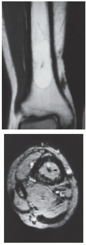
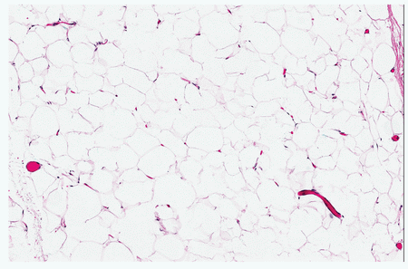
A) Intramedullary bone infarction.
B) Liposarcoma of bone.
C) Intraosseous lipoma.
D) Intraosseous ganglion.
E) Langerhans cell histiocytosis.
View Answer
Answer to question 65: C
Coronal T1-weighted MR image shows a well-demarcated lesion exhibiting high signal intensity paralleling the signal of subcutaneous fat, with internal focus of low signal intensity. Axial T2-weighted MRI demonstrates that the lesion now becomes of intermediate signal, again paralleling the signal intensity of subcutaneous fat. The focus of low signal intensity (arrowhead) represents a calcification. These are the characteristic imaging features of intraosseous lipoma. The diagnosis is confirmed on the histopathologic section that shows benign mature lipocytes possessing a large clear cytoplasmic vacuoles that displace the crescent-shaped nuclei to the periphery. There is no evidence of lipoblasts or atypical stellate spindle cells with mitotic figures that are seen in malignant lipomatous tumors. (A) Intramedullary bone infarct may contain fatty tissue; however, it has characteristic MRI appearance. On T1-weighted images, there is a serpentine band of low signal intensity representing the reactive interface surrounding the central area of bone necrosis; T2-weighted and STIR images demonstrate serpentine low signal line adjacent to a high signal intensity line (so-called double-line sign). (B) Liposarcoma of bone shows more aggressive imaging features than intraosseous lipoma, and histopathology confirms malignancy demonstrating lipoblasts, atypical stellate, and spindle cells in myxoid stroma, as well as prominent pleomorphism and numerous mitotic figures. (D) Intraosseous ganglion, most of the time exhibits a sclerotic border, shows no internal calcifications or ossifications, and MRI features of intermediate signal intensity on T1-weighted sequences and high signal on T2 weighting are not consistent with a fatty tumor, but rather with a cystic-like lesion. (E) Langerhans cell histiocytosis demonstrates no fat signal on any of MRI sequences, and shows characteristic histopathologic features that include aggregates of histiocyticappearing cells in a mixed inflammatory background with prominent eosinophilia, and Langerhans cells with indistinct cytoplasmic borders and kidney-shaped grooved nuclei.
Question 66
Regarding liposarcoma of bone, which one of the following statements is FALSE?
A) Presents as a painful mass.
B) Histopathologic variants include well-differentiated, myxoid, and pleomorphic.
C) Majority of the patients are children.
D) It is a tumor of long tubular bones.
E) Behavior of the tumor correlates with histopathologic grade.
View Answer
Answer to question 66: C
Liposarcoma of bone occurs in all age groups, but the majority of the patients are adults. The remaining statements are all true.
Question 67
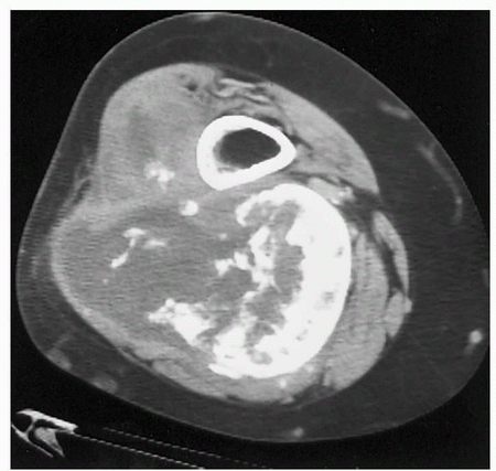
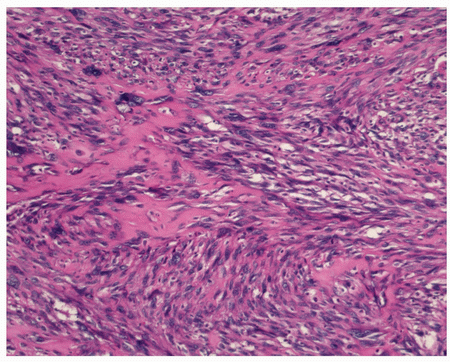
A 68-year-old woman presented with a progressively enlarging soft-tissue mass in the popliteal region of her right knee. Based on CT and histopathologic section, what is the most likely diagnosis?
 |
 |
A) Myositis ossificans.
B) Periosteal chondroma.
C) Soft-tissue chondrosarcoma.
D) Soft-tissue osteosarcoma.
E) None of the above.
View Answer
Answer to question 67: D
CT section shows a large soft-tissue mass adjacent to the posterior cortex of the distal femur exhibiting ossifications, some well organized, particularly at the periphery (right side), some scattered centrally (left side) with reverse zoning pattern. The cortex of the bone is not invaded. These imaging features are characteristic of soft-tissue (extraskeletal) osteosarcoma. The diagnosis is confirmed by histopathologic section that demonstrates cell-rich malignant tumor with fibrosarcomatous areas and tumor osteoid formation by malignant cells. (A) Imaging features of myositis ossificans may resemble soft-tissue osteosarcoma, the ossifications, however, instead of being centrally scattered within the mass, are well organized at the periphery of the lesion, creating so-called zonal phenomenon. (B) Lesion of periosteal chondroma, instead of ossifications, contains chondroid calcifications. In addition, it commonly erodes the adjacent cortex, forming buttress of periosteal reaction. Histopathology shows hyaline cartilage formed by chondrocytes and absence of tumor osteoid. (C) Soft-tissue chondrosarcoma may mimic soft-tissue osteosarcoma; however, instead of ossifications, it contains calcifications. On histopathologic examination, the tumor is characterized by undifferentiated mesenchymal cells with only rare islands of well-differentiated cartilage and may also exhibit myxoid changes.
Question 68
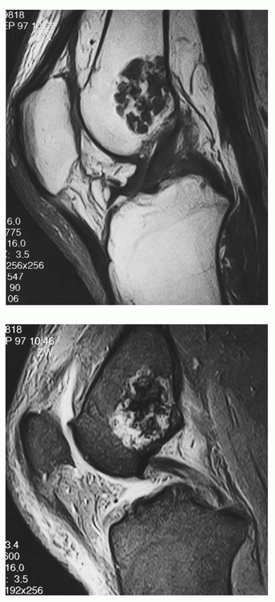
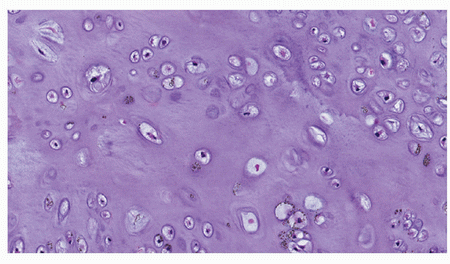
A) Enchondroma.
B) Low-grade chondrosarcoma.
C) Chondromyxoid fibroma.
D) Periosteal chondroma.
E) Chondroblastoma.
View Answer
Answer to question 68: A
MRI examination shows a classic appearance of enchondroma. Coronal T1-weighted MR image of the knee shows a well-defined lobulated lesion in the distal femur exhibiting intermediate-to-low signal intensity. Centrally located foci of low signal represent calcifications. Coronal T2-weighted MRI shows the lesion becomes of high signal, with calcifications remaining of low signal intensity. Histopathologic section confirms the classic features of enchondroma: sparsely cellular tissue and uniform-sized chondrocytes located in rounded lacunae. (B) Imaging features of low-grade chondrosarcoma may resemble an enchondroma, but cortex is usually focally thickened, and there is evidence of deep endosteal scalloping of the cortex. Histopathology shows cellular pleomorphism and hyperchromatism of the nuclei. (C) Chondromyxoid fibroma shows slightly more aggressive imaging features, periosteal reaction, and conspicuous lack of calcifications. On histopathologic examination, chondromyxoid fibroma is characterized by large lobulated areas of spindle-shaped or stellate cells distributed within abundant myxoid and chondroid intercellular matrix. (D) Periosteal chondroma is a surface, not medullary tumor. Histopathology is very similar to enchondroma, although the tumor tissue is more cellular, and some cells may appear atypical. (E) Chondroblastoma is an epiphyseal lesion, commonly seen in the immature skeleton. MRI shows usually heterogeneous signal of the tumor. Histopathologic features are characteristic, including nodules of fairly mature cartilaginous matrix surrounded by a highly cellular, relatively undifferentiated tissue composed of round and polygonal chondroblast-like cells and giant cells. In addition, the almost pathognomonic fine lattice-like matrix calcifications are present having a spatial arrangement that resembles the hexagonal configuration of chicken wire.
Question 69
Regarding chondroblastoma, all of the following statements are true, EXCEPT for which one?
A) Imaging studies may resemble features of clear cell chondrosarcoma.
B) It is an epiphyseal lesion.
C) It may metastasize to the lungs.
D) Most tumors are DNA diploid.
E) Osteoclast-like giant cells are not a part of histopathologic spectrum.
View Answer
Answer to question 69: E
Giant cells of the osteoclast-like or chondroclast type are usually part of the histopathologic spectrum of chondroblastoma. The remaining statements are all true.
Question 70
Regarding periosteal chondroma, which of the following statements is NOT true?
A) Preferential sites include the proximal humerus, femur, and tibia.
B) Unlike an enchondroma, majority of the cases are seen in the sixth and seventh decades of life.
C) Bone erosion is commonly present.
D) The tumor tissue is more cellular than enchondroma.
E) Imaging features of some lesions may mimic osteochondromas and aneurysmal bone cysts.
View Answer
Answer to question 70: B
Periosteal chondroma occurs in children and young adults, most cases seen in the second and third decades of life. The remaining statements are all true.
Question 71
Concerning telangiectatic osteosarcoma, which of the following statements is (are) true?
A) It represents one of the most aggressive type of osteosarcomas.
B) Most of these tumors arise in the femur or tibia.
C) Imaging and histopathologic features may resemble those of aneurysmal bone cyst.
D) A and C only.
E) All statements are correct.
View Answer
Answer to question 71: E
(A) Telangiectatic osteosarcoma represents one of the most aggressive types of osteosarcomas. It affects males twice as often as females and occurs predominantly during the second and third decades of life. Originally described by Paget in 1854 and subsequently redefined by Gaylord in 1903 as a “malignant bone aneurysm,” it was renamed as “hemorrhagic osteosarcoma” by Campanacci in 1971. (B) Most of the tumors arise in the femur and tibia. (C) Imaging features of telangiectatic osteosarcoma and aneurysmal bone cyst may overlap, particularly both lesions may exhibit soft-tissue mass and fluid-fluid levels on MRI. Likewise, the histopathologic features of both lesions overlap. In fact, the confusion in diagnosis between telangiectatic osteosarcoma and aneurysmal bone cyst represents one of the most treacherous pitfalls in tumor pathology. The hallmark of telangiectatic osteosarcoma is the presence of anaplastic cells with atypical mitoses forming tumor bone or tumor osteoid, the feature not present in aneurysmal bone cyst. However, some of these tumors exhibit only minimal amount of osteoid production, making it difficult to find it on the biopsy specimens.
Question 72
A 14-year-old boy on MRI studies was diagnosed as having a bone tumor. Based on the histopathologic section, what is the most likely diagnosis?
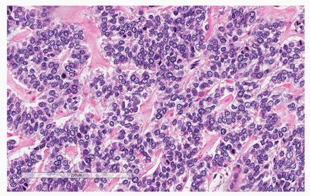 |
A) Osteoblastoma.
B) Fibrous dysplasia.
C) Small cell osteosarcoma.
D) Telangiectatic osteosarcoma.
E) Periosteal osteosarcoma.
View Answer
Answer to question 72: C
The histopathologic section shows malignant small round and oval cells producing tumor osteoid (pink material); therefore, the most likely diagnosis is small cell osteosarcoma. Some other malignant and benign lesions may exhibit small round cells, such as Ewing sarcoma, lymphoma, myeloma, mesenchymal chondrosarcoma, metastatic neuroblastoma, or Langerhans cell histiocytosis. Contrary to the small cell osteosarcoma, however, neither of the listed tumors is able to produce tumor osteoid, the hallmark of osteosarcomas. (A) Cells of osteoblastoma are able to produce osteoid but not malignant variety. (B) Fibrous dysplasia is characterized by metaplastic immature woven bone trabeculae without osteoblasts, haphazardly arranged in the fibrous stroma. (D) Telangiectatic osteosarcoma rarely shows tumor osteoid production, and the cells are not small and round, but rather fusiform and spindle. In addition, the giant cells are commonly present in the field of view. (E) Periosteal osteosarcoma shows predominantly cartilaginous component, and the tumor cells, particularly at the periphery, are not round but spindled.
Question 73
A 55-year-old woman presented with left knee pain after a fall. The radiograph was obtained followed by CT. The radionuclide bone scan (not shown here) demonstrated a moderate uptake of the radiopharmaceutical tracer. What should be done next?
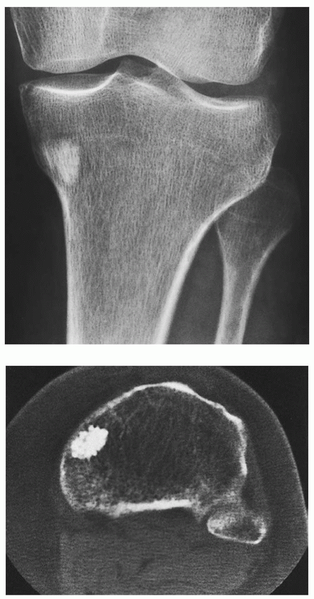 |
A) Perform MRI for better characterization of the lesion.
B) The lesion looks like osteosarcoma; therefore, an open biopsy should be performed.
C) The lesion looks like a metastatic tumor; therefore, an extensive clinical workup has to be done to look for the primary source.
D) The lesion looks like an osteoma; therefore, a clinical workup should be done to exclude Gardner syndrome.
E) The lesion shows characteristic features of a bone island; therefore, nothing invasive should be contemplated; however, to repeat radiographs in about 6 months is a prudent action.
View Answer
Answer to question 73: E
Anteroposterior radiograph of the knee shows a homogeneously sclerotic lesion with small radiating streaks (thorny radiations) in the proximal tibia. CT section shows a highattenuation lesion with a rough border. These are the characteristic imaging features of a bone island (enostosis). Some of the bone islands may show positive radionuclide bone scan due to increased metabolism and osteoblastic activity. The best action is to follow-up the patient with repeat imaging studies in about 6 months. (A) The radiographic and CT features of bone island including homogeneity of the lesion and rough border (pseudopodia, thorny radiations, radiating streaks) are diagnostic, and there is no need for additional imaging studies. (B) The lesion definitely does not exhibit imaging features of osteosarcoma because there is lack of aggressive findings, and furthermore, osteosarcoma most commonly occurs in children and adolescents. (C) Metastasis always should be considered in patients older than 50 years. However, imaging features, particularly the rough border of the lesion and only moderate uptake of the tracer on scintigraphy is more consistent with a bone island. (D) Osteoma is a surface and not an intramedullary lesion, so this diagnosis can be assertively excluded.
Question 74
Which of the following lesions SHOULD NOT be biopsied? Choose all that are pertinent.
A) 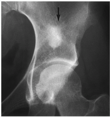
B) 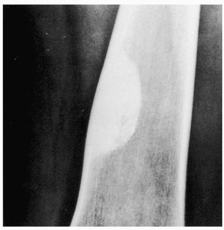
C) 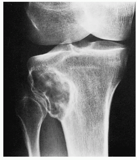
D) 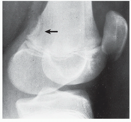
E) 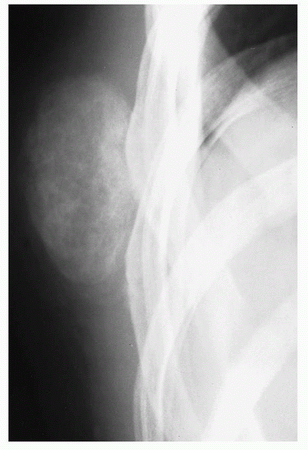
View Answer
Answer to question 74: A,B,D,E
The only lesion that should undergo biopsy is the one shown in part (C). The radiograph of the knee shows an eccentric radiolucent lesion with sclerotic border and central calcifications in the lateral aspect of the proximal tibia. The lesion exhibits nonaggressive features including a narrow zone of transition and most likely represents an enchondroma, focus of fibrous dysplasia, nonossifying fibroma, or benign fibrous histiocytoma. The possibility of chondrosarcoma, however, cannot be completely excluded, since the low-grade cartilage tumors may masquerade as benign lesions. Biopsy of the lesion is a prudent action. To the contrary, all other lesions under consideration have a very characteristic radiographic appearance, and diagnosis can be made with high degree of accuracy without the biopsy. These conditions belong to the group of so-called don’t touch lesions. (A) The radiograph of the left hip shows a large sclerotic lesion in the ilium exhibiting rough border and “thorny radiations” merging with the normal trabeculae of the host bone (arrow). Those are the characteristic imaging features of bone island (enostosis). The proper action is not to biopsy the lesion but to follow-up the patient with repeat radiologic studies in about 6 months. (B) The radiograph of the femur shows a homogeneously sclerotic lesion abutting the medial cortex with narrow zone of transition. The cortex is normal in thickness, and there is no evidence of a soft-tissue mass. These are the characteristic imaging features of healed nonossifying fibroma. No further action is required. (D) An oblique radiograph of the knee in a child (observe the open growth plate) shows a saucer-like defect in the medial cortex of the distal femoral metaphysis at the linea aspera, producing cortical irregularity (arrow). This is a classic presentation of benign periosteal desmoid. It is a tumor-like proliferation of the periosteum, which has a striking predilection for the posteromedial cortex of the medial femoral condyle. The lesion usually occurs between the ages 12 and 20 years, mainly in boys, and most lesions disappear spontaneously by the end of the second decade. Periosteal desmoid is considered to represent asymptomatic normal variant that does not require biopsy or treatment. (E) A coned-down radiograph of the right ribs shows a large ossific mass in the soft tissues. There is increased density on the periphery of the lesion and more radiolucent center, referred to as zoning phenomenon. In addition, a narrow radiolucent cleft separates the mass from the ribs. These are the characteristic imaging features of juxtacortical myositis ossificans. The zoning phenomenon represents the maturation pattern of the lesion, which is undifferentiated and cellular in the center, and exhibits an increasingly mature ossification toward the periphery. With time, the lesion decreases in size and eventually may disappear completely, or persists as a well-matured ossific focus. It should never be biopsied, although timely repeated imaging studies to document regression of the lesion is a proper action.
Question 75
Which one of the following conditions is responsible for most osteosarcomas seen in patients older than 50 years?
A) Fibrous dysplasia.
B) Prior radiation.
C) Paget disease.
D) Chronic draining sinus tract of osteomyelitis.
E) Intramedullary bone infarct.
View Answer
Answer to question 75: C
A large number of secondary osteosarcomas arise as a complication of Paget disease and characteristically develop in pagetic bone. Most cases of malignant transformation occur in polyostotic Paget disease. Osteosarcoma can develop in any affected bone, although the pelvic bones and femur are the preferential sites of this complication. The other sites commonly affected are the humerus and the tibia. Patients with advanced Paget disease are at greater risk. (A) Osteosarcoma arising in fibrous dysplasia is a rare event occurring in about 0.5% to 1% of patients with this disorder. In reported cases, the majority of the patients had a polyostotic fibrous dysplasia. The affected sites included craniofacial bones, proximal femur, humerus, and pelvis. (B) Radiation therapy for an unrelated previous conditions may also give rise to secondary osteosarcoma. These malignancies account for 3% to 5% of all osteosarcomas, and 50% to 60% of radiation-induced sarcomas. The threshold is approximately 800 to 1,000 cGy, but usually, a dose of at least 3,000 cGy administered within 3 weeks is required to develop this complication. The latent period is between 4 and 42 years, with an average of 11 years. Postirradiation osteosarcoma may develop either after radiation therapy for benign bone lesions, such as fibrous dysplasia and giant cell tumor, or after irradiation of malignant conditions such as breast cancer and lymphoma. These tumors may also develop as a result of ingestion and intraosseous accumulation of radioisotopes, as has been described in painters of radium watch dials. (D) One of the most common complications of chronic draining sinus tract of osteomyelitis is development of squamous cell carcinoma. Less commonly fibrosarcoma and osteosarcoma may develop. In most patients with osteomyelitis, the history of the disease dates to childhood, and sinuses draining for more than 20 years are generally the precursors of malignant neoplasms. (E) Secondary osteosarcoma associated with medullary bone infarcts is extremely rare complication. More commonly reported malignancies arising in bone infarct are fibrosarcoma, malignant fibrous histiocytoma, and angiosarcoma.
ANSWERS
Answer to question 1: A
The radiograph shows a radiolucent lesion (arrow) within the medial femoral cortex exhibiting narrow zone of transition and surrounded by sclerotic area. There is associated well-organized periosteal reaction. Histopathologic section shows sharply circumscribed lesion containing bone trabeculae within vascularized connective tissue, lined by the plump osteoblasts and surrounded by sclerotic bone. Both, imaging characteristics and histopathologic features, are consistent with osteoid osteoma. (B) Chondroblastoma usually is present in the immature skeleton and is invariably located in the epiphysis. Histopathology shows chondroid matrix and not osteoblasts. (C) Osteofibrous dysplasia predominantly occurs in childhood (more than 60% of the patients are younger than 12 years old) and affects the cortex of the tibia (not femur). Histopathology shows characteristic immature woven bone trabeculae with peripherally displayed lamellar bone, lined by osteoblasts (“dressed trabeculae”). (D) Osteosarcoma and (E) Ewing sarcoma are predominantly intramedullary tumors, exhibiting more aggressive imaging characteristics. Histopathology shows malignant cells.
Answer to question 2: C
Maffucci syndrome is characterized by multiple enchondromas (Ollier disease) and cavernous hemangiomas located in the skin and soft tissues. This syndrome carries a greater risk of chondrosarcoma as well as nonskeletal malignancies, particularly of the ovaries and brain. (A) von Recklinghausen disease of bone, also known as osteitis fibrosa cystica generalisata, refers to severe bone disorder of primary hyperparathyroidism, characterized by increased osteoblastic activity, osteoclastic resorption, peritrabecular fibrosis, and brown tumors. (B) Li-Fraumeni syndrome caused by inherited mutation of p53 is associated with greater risk of sarcomas (particularly osteosarcoma) and other malignancies, such as breast cancer, brain tumors, and leukemias. (D) Hereditary multiple exostoses, also known as diaphyseal aclasis, is an autosomal dominant disorder characterized by the presence of multiple osteochondromas, particularly at the knee, hip, ankle, and shoulder joints. (E) Gardner syndrome, an autosomal dominant disorder, consists of a combination of multiple osteomas, intestinal polyposis, skin fibromatosis, and epidermal sebaceous cysts.
Answer to question 3: A
Mutation of p53 (Li-Fraumeni syndrome) is associated with greater risk of sarcomas, particularly 500-fold greater risk of osteosarcoma. (B) Translocation t(X;18) is characteristic of synovial sarcoma. (C) Translocation t(12;16) is characteristic of myxoid liposarcoma. (D) 1p rearrangements are characteristic for conventional chondrosarcoma. (E) Translocation t(9;22) is characteristic for myxoid chondrosarcoma.
Answer to question 4: D
(A) Lymphoma, (B) metastatic neuroblastoma, (C) mesenchymal chondrosarcoma, and (E) embryonal rhabdomyosarcoma show similar morphologic features to Ewing sarcoma, all characterized by small round blue tumor cells. Leiomyosarcoma of bone, on the other hand, is composed of bundles of fascicles of spindle cells.
Answer to question 5: C
Parosteal osteosarcoma is low-grade (grade 1) sarcoma. (B) Periosteal osteosarcoma is a grade 2, and (A) Paget osteosarcoma, (D) telangiectatic osteosarcoma, and (E) small cell osteosarcoma are high-grade (grade 3) sarcomas.
Answer to question 6: B
The epithelial component of adamantinoma expresses positivity for cytokeratins 14 and 19. CK8 and CK18 are expressed in epithelioid component of synovial sarcoma. The statements (A), (C), (D), and (E) are all correct.
Answer to question 7: B
The radiograph shows well-defined eccentric radiolucent lesion with sclerotic lobulated border, exhibiting no evidence of periosteal reaction (arrow), all imaging features of nonossifying fibroma. Histopathology shows fibrous stroma displaying a storiform arrangement, and several scattered giant cells, confirming the radiologic diagnosis. (A) Giant cell tumor almost always occurs in skeletally mature patients and extends into the articular end of bone. Generally, it does not exhibit a sclerotic border. Histopathology shows mononuclear cells intermixed with numerous uniformly distributed giant cells. (C) Aneurysmal bone cyst characteristically shows expansive features and well-organized periosteal reaction in the form of a buttress. Histopathology shows multiple blood-filled spaces alternating with more solid areas. (D) Chondroblastoma is an epiphyseal lesion, exhibiting chondroid calcifications. Histopathology shows nodules of fairly mature cartilaginous matrix surrounded by a highly cellular tissue composed of round and polygonal chondroblast-like cells, and matrix calcifications having a spatial arrangement that resembles the hexagonal configuration of chicken wire. (E) Giant cell-rich osteosarcoma exhibits aggressive imaging features with poorly defined borders showing wide zone of transition. Histopathology is similar to that of a giant cell tumor; however, tumor bone or tumor osteoid is present in the vicinity of malignant cells.
Answer to question 8: B
The radiograph shows an eccentric radiolucent lesion with narrow zone of transition and geographic type of bone destruction, exhibiting a noninterrupted periosteal reaction in form of a buttress, all typical imaging features of
aneurysmal bone cyst (ABC). Histopathology shows plump fibroblasts and giant cells aggregating around and within the blood-filled spaces, some of them showing characteristic feature of “jumping into the swimming pool”, confirming the radiologic diagnosis. (A) Nonossifying fibroma exhibits no expansive imaging features and lack of periosteal reaction. Histopathology shows predominantly bland monomorphous spindle cells admixed with histiocytic cells frequently arranged in a swirling storiform pattern and occasionally scattered giant cells. (C) Simple bone cyst shows almost invariably central location and lack of periosteal reaction, unless there is a pathologic fracture present. Histopathology shows walls of the cyst covered by a flattened single-cell lining. (D) Giant cell tumor, although occasionally may exhibit expansive imaging features, generally shows lack of sclerotic border and lack of buttress type of periosteal reaction, characteristic features of ABC. Histopathology shows mononuclear cells intermixed with numerous uniformly distributed giant cells. (E) Telangiectatic osteosarcoma, although occasionally similar in appearance to ABC, shows more aggressive imaging features associated with a soft-tissue mass. Interrupted periosteal reaction in form of lamellar, sunburst, or Codman triangle is commonly present. Histopathology shows malignant cells forming tumor bone or tumor osteoid.
aneurysmal bone cyst (ABC). Histopathology shows plump fibroblasts and giant cells aggregating around and within the blood-filled spaces, some of them showing characteristic feature of “jumping into the swimming pool”, confirming the radiologic diagnosis. (A) Nonossifying fibroma exhibits no expansive imaging features and lack of periosteal reaction. Histopathology shows predominantly bland monomorphous spindle cells admixed with histiocytic cells frequently arranged in a swirling storiform pattern and occasionally scattered giant cells. (C) Simple bone cyst shows almost invariably central location and lack of periosteal reaction, unless there is a pathologic fracture present. Histopathology shows walls of the cyst covered by a flattened single-cell lining. (D) Giant cell tumor, although occasionally may exhibit expansive imaging features, generally shows lack of sclerotic border and lack of buttress type of periosteal reaction, characteristic features of ABC. Histopathology shows mononuclear cells intermixed with numerous uniformly distributed giant cells. (E) Telangiectatic osteosarcoma, although occasionally similar in appearance to ABC, shows more aggressive imaging features associated with a soft-tissue mass. Interrupted periosteal reaction in form of lamellar, sunburst, or Codman triangle is commonly present. Histopathology shows malignant cells forming tumor bone or tumor osteoid.
Answer to question 9: D
The radiograph shows a radiolucent lesion with fine matrix calcifications, exhibiting a narrow zone of transition and thin sclerotic border, located in the epiphysis of the proximal tibia and abutting the growth plate, all characteristic features of chondroblastoma. Histopathology showing polygonal chondroblasts with indistinct outlines and uniformly shaped nuclei, scattered giant cells, and fine “chicken-wire” calcifications confirm this diagnosis. (A) Giant cell tumor, although also occurring in the end of bone, invariably is seen in the mature skeleton, after the growth plate has been obliterated. In addition, it never exhibits matrix mineralization. Histopathology shows numerous large giant cells uniformly distributed throughout the lesion. (B) Nonossifying fibroma is a diaphyseal lesion, never occurring in the epiphysis and shows no evidence of matrix calcifications. Histopathology shows whorled pattern of fibrous tissue containing giant cells, hemosiderin, and lipid-laden histiocytes. (C) Osteosarcoma generally shows a metaphyseal or diaphyseal location and exhibits more aggressive imaging features commonly with sclerotic foci representing tumor-bone formation. Soft-tissue mass is a typical feature. Histopathology shows malignant cells forming tumor osteoid and tumor bone and invasion of marrow spaces and bone trabeculae. (E) Aneurysmal bone cyst is located in the diaphysis or metaphysis, show blown-up imaging features, and lack of mineralization. It also exhibits a well-organized periosteal reaction. Histopathology shows blood-filled vascular spaces bordered by solid areas containing spindle cells and giant cells with occasional foci of osteoid.
Answer to question 10: D
Malignant transformation of a solitary osteochondroma is extremely rare and was reported in less than 1% of cases. The statements (A), (B), (C), and (E) are all correct.
Answer to question 11: D
Giant cell tumor commonly exhibits lack of sclerotic border. The statements (A), (B), (C), and (E) are all correct.
Answer to question 12: B
MR images show a signal abnormality in the right proximal femur extending into the middle of the shaft. In the intertrochanteric region, there are at least two lesions with sclerotic border. In the soft tissue, there is a well-circumscribed lesion showing homogeneous high signal intensity on T2-weighted sequences and rim enhancement on the postcontrast study. All lesions show absence of aggressive features. These findings combined with the clinical history of long-standing deformity are consistent with polyostotic fibrous dysplasia and soft-tissue myxoma, features of Mazabraud syndrome. (A) Osteosarcoma will show more aggressive imaging features, and it extremely rare metastasizes to the soft tissue. Besides, metastatic soft-tissue lesion will not exhibit homogeneous high signal on T2 weighting, but rather heterogeneity with some evidence of bone formation. (C) Jaffe-Campanacci syndrome consists of coexistence of nonossifying fibromas and neurofibromatosis, associated with formation of caféau-lait spots on the skin. (D) Rothmund-Thomson syndrome, also known as congenital poikiloderma, is a hereditary disorder characterized by erythematous and maculopapular skin lesions with areas of hyperpigmentation and coexistence of conventional osteosarcoma. (E) Werner syndrome, also known as adult progeria, is characterized by premature aging, scleroderma-like skin changes, and coexistence of osteosarcomas.
Answer to question 13: C
Radiograph shows a several small radiolucent channels traversing the medullary portion and the cortex of the proximal phalanx of the middle finger associated with a soft-tissue prominence. Coronal T2-weighted fat-suppressed MRI shows high signal intensity lesion affecting both the bone and soft tissue. Low signal intensity foci represent the phleboliths. From the given choices, the most likely diagnosis is an intraosseous hemangioma with soft-tissue extension. (A) Ewing sarcoma is a malignant tumor predominantly affecting the long and flat bones, and involvement of the short tubular bone, although reported in the literature, is extremely rare. (B) The possibility of angiosarcoma should be considered in the differential diagnosis; however, majority of the reported cases were affecting long bones and bones of the pelvis. Involvement of the short tubular bone is unlikely. In addition, the imaging features of angiosarcoma are more aggressive than presented in this case. (D) Langerhans cell histiocytosis is a condition predominantly affecting children, with peak incidence between the ages of 5 and 10 years.
The sites of involvement include the axial skeleton and the long bones. Periosteal reaction is usually lamellated. Phleboliths are not the part of presentation. (E) Although the short tubular bones are the common sites of enchondroma, this benign cartilaginous lesion does not extend into the soft tissues, unless it is multifocal such as in Ollier disease.
The sites of involvement include the axial skeleton and the long bones. Periosteal reaction is usually lamellated. Phleboliths are not the part of presentation. (E) Although the short tubular bones are the common sites of enchondroma, this benign cartilaginous lesion does not extend into the soft tissues, unless it is multifocal such as in Ollier disease.
Answer to question 14: E
Lateral radiograph of the skull shows numerous variousin-size punched-out lesions, characteristic of multiple myeloma. CT section shows predominant involvement of the diploë, also typical feature for myeloma. (A) Osteolytic metastases to the skull commonly present as a larger areas of destruction. In contrast to myeloma, the inner and outer tables are also affected. (B) Skull abnormalities seen in hyperparathyroidism consist of osteopenia, which gives a characteristic granular appearance of the bones of the vault, referred to as “salt and pepper” appearance, not the punchedout lesions. (C) Fibrous dysplasia affects predominantly the base of the skull in form of areas of sclerosis. (D) Osteolytic phase of Paget disease in the skull is known as osteoporosis circumscripta, appearing as a large area of geographic bone destruction. Mixed and cool phases of the disease affecting the skull manifest as areas of ill-defined focal densities resembling cotton balls.
Answer to question 15: A
One of the characteristic features of multiple myeloma, in contrast to metastatic disease, is essentially normal radionuclide bone scan. The remaining statements (B), (C), (D), and (E) are all correct.
Answer to question 16: B
One of the characteristic features of polyostotic fibrous dysplasia affecting the femur is so-called shepherd’s crook deformity, which is commonly the result of multiple pathologic fractures. (A) Histopathology of fibrous dysplasia shows immature woven bone trabeculae without osteoblastic rimming, so-called naked trabeculae. (C) Skull lesions in fibrous dysplasia are seen predominantly at the base of the skull in form of increased sclerosis. (D) Postcontrast MRI shows invariably enhancement of the lesions of fibrous dysplasia. (E) Gains of chromosomes 1q, 3q, 9q, 11q, and 15q are characteristic features of multiple myeloma, not polyostotic fibrous dysplasia. McCune-Albright syndrome, which is the association of polyostotic fibrous dysplasia with endocrine disturbances (such as premature sexual development, gigantism or acromegaly, hyperparathyroidism, and Cushing syndrome) and skin pigmentation (cafe-au-lait spots), is caused by somatic mutations in the GNASI gene located in chromosome 20q13.
Answer to question 17: C
The radiograph of the hand shows multiple sharply marginated radiolucent lesions within the metacarpals and phalanges, some within the cortex, typical for enchondromas. In addition, there is growth stunting of the distal ulna. The combination of these findings is characteristic for Ollier disease. (A) Polyostotic fibrous dysplasia exhibits thinning of the cortex associated with smoky or ground-glass appearance of the medullary portion of bones, without sharply marginated radiolucent lesions typical for enchondromatosis. In addition, overgrowth of the bone rather than growth stunting is occasionally present. (B) The most important identifying features of osteochondromas are that the cortex of the lesion merges without interruption with the cortex of the host bone and that the cancellous portion of the lesion is continuous with the medullary cavity of the adjacent diaphysis, findings not present in the case under consideration. (D) Maffucci syndrome consists of association of Ollier disease with softtissue hemangiomas. The radiography will invariably demonstrate phleboliths, which appear as round calcific foci, not present on the submitted radiograph. (E) Neurofibromatosis may be associated with osseous deformities, characteristic pit-like cortical erosions, and soft-tissue masses, features not present in the case under consideration.
Answer to question 18: B
Anteroposterior and lateral radiographs show ill-defined sclerotic lesion in the medullary portion of the left distal femur associated with a large, dense soft-tissue mass. Sagittal T1-weighted fat-suppressed MR image obtained after intravenous administration of gadolinium shows significant enhancement of both medullary tumor and soft-tissue mass. Nonenhanced area represents tumor bone formation. The imaging features are typical for conventional osteosarcoma. (A) Chondrosarcoma, although may present as a sclerotic lesion, most of the time exhibits characteristic chondroid calcifications in the form of stippling, flocculent densities, or rings and arcs (“o”s and “c”s). In addition, there is invariably present a deep endosteal scalloping and cortical thickening, absent in the case under consideration. (C) Parosteal osteosarcoma is a surface lesion, usually exhibiting well-defined sclerotic mass outside of the cortex, which is slightly less dense on the periphery. It may possess an incomplete cleft between the tumor and underlying cortex. (D) Periosteal osteosarcoma is also a surface lesion, displaying ill-defined heterogeneous ossifications with calcified spiculations interspersed with areas of radiolucencies representing uncalcified matrix. (E) Melorheostosis, a form of sclerosing dysplasia, may occasionally mimic osteosarcoma, although the surface ossifications, in contrast to osteosarcoma, are well defined, resembling wax dripping on one side of the candle.
Answer to question 19: B
Histopathologic section shows typical appearance of synovial (osteo)chondromatosis. Several cartilaginous nodules are seen covered by a thin layer of synovium. Although the nodules are highly cellular, there is no evidence of spindle cells. All other statements (A), (C), (D), and (E) are correct.
Answer to question 20: A
Histopathologic section shows the classic features of the giant cell tumor: fibrocytic and monocytic stromal cells and large giant cells containing numerous nuclei. The preferential location of this tumor is the articular end of bone in skeletally mature patient and not in the metaphysis. The remaining statements (B), (C), (D), and (E) are all correct.
Answer to question 21: C
Jaffe-Campanacci syndrome consists of association of multiple nonossifying fibromas with neurofibromatosis.
Answer to question 22: E
Osteofibrous dysplasia, also known as Kempson-Campanacci lesion, characteristically affects children, with about 60% of the affected patients being younger than 5 years. The preferential site is the anterior cortex of the tibia.
Answer to question 23: C
Mesenchymal chondrosarcoma is an aggressive tumor with common distant metastases. The remaining statements (A), (B), (D), and (E) are all correct.
Answer to question 24: E
All statements concerning clear cell chondrosarcoma are true.
Answer to question 25: B
Histopathologically, osteoid osteoma and osteoblastoma exhibit similar features. Both lesions show irregular woven bone trabeculae with various degrees of mineralization, osteoblasts and giant cells, ectatic blood vessels, and reactive periosteal shell of new bone. The difference is in size: osteoid osteoma exhibits limited growth potential and size less than 2 cm, whereas osteoblastoma is commonly greater than 2 cm in size.
Answer to question 26: D
Anteroposterior radiograph of the right knee shows fluffy sclerotic densities in the metaphysis and proximal diaphysis of the tibia representing tumor bone formation, associated with velvet-type periosteal reaction. Similar densities are seen within the soft-tissue mass at the site of the proximal fibula. These are the typical imaging features of conventional osteosarcoma. The diagnosis is confirmed by the histopathologic section, showing markedly pleomorphic osteoblastic tumor cells with hyperchromatic nuclei forming lace-like osteoid. (A) Imaging features of osteoblastoma occasionally may mimic those of osteosarcoma; however, the lesion exhibits more focally confined densities, and the periosteal reaction is solid, not interrupted as in the case under consideration. Histopathology shows large but benign osteoblasts forming immature woven bone trabeculae. (B) Osteoid osteoma shows resemblance to osteoblastoma but exhibits completely different imaging features than osteosarcoma. The size of the lesion is usually less than 2 cm, and periosteal reaction is of solid type. Moreover, the lesion does not produce the soft-tissue mass. Histopathology shows a well-defined nidus composed of osteoid and trabeculae of immature woven bone, surrounded by sclerotic reactive bone. (C) Imaging features of giant cell-rich osteosarcoma may be indistinguishable from that of conventional osteosarcoma; however, histopathology shows striking resemblance to giant cell tumor with numerous osteoclast-like giant cells and scant osteoid formation, unlike the histopathologic section under consideration. (E) Callus formation of healing fracture shows more organized pattern on imaging studies, and there is no softtissue mass present. Furthermore, histopathology shows lack of malignant cells and lack of tumor bone or tumor osteoid.
Answer to question 27: D
Lipoma arborescent is a rare intra-articular disorder characterized by a nonneoplastic lipomatous proliferation of the synovium resembling a frond-like mass. It is more prevalent in males and usually diagnosed in the fourth and fifth decades of life. MRI features are characteristic and diagnostic for this disorder, showing fat signal intensity on all imaging sequences.
Answer to question 28: E
Synovial (osteo)chondromatosis is marked by the metaplastic proliferation of multiple cartilaginous nodules in the synovial membrane of the joints, bursae, or tendon sheaths. The knee joint is a preferential site of involvement. Joint effusion, tenderness, limited motion in the joint, and a softtissue mass are common clinical findings. Histopathology shows numerous highly cellular cartilaginous nodules. The cells with plump and dark nuclei are arranged in clusters and show moderate pleomorphism.
Answer to question 29: B
Lateral radiograph of the distal femur shows intramedullary lesion with wide zone of transition and chondroid calcifications. In addition, noted is focal cortical thickening and periosteal reaction (arrows). These findings are also demonstrated on the axial CT section through the tumor. The histopathology shows pleomorphic tumor with hyperchromatic nuclei permeating the bone trabeculae. The imaging findings and histopathology are characteristic for conventional intramedullary chondrosarcoma. (A) Although enchondroma also may display chondroid calcifications, generally there is no periosteal reaction, unless the pathologic fracture has occurred. Also, the cortex is not thickened; to the contrary, most of the time noted is thinning of the cortex. (C) Medullary bone infarction also exhibits calcifications, but in contrast to the chondroid tumors, the calcifications are coarser, and peripheral serpentine sclerotic fibro-osseous wall is commonly observed. Moreover, the cortex is not thickened. (D) Chondromyxoid fibroma exhibits narrow zone of transition and lack of visible calcifications. (E) Chondroblastoma is
a tumor of immature skeleton, invariably located within an epiphysis. It exhibits narrow zone of transition and lack of periosteal reaction at the site of the lesion.
a tumor of immature skeleton, invariably located within an epiphysis. It exhibits narrow zone of transition and lack of periosteal reaction at the site of the lesion.
Answer to question 30: A
Chondromyxoid fibroma is the only cartilaginous lesion that does not show visible calcifications on radiography. The remaining statements (B), (C), (D), and (E) are all correct.
Answer to question 31: B
The histopathologic section shows uniformly arranged small round cells exhibiting indistinguished borders, with darkly stained round nuclei, and scanty eosinophilic cytoplasm, highly characteristic for Ewing sarcoma. (A) Langerhans cell histiocytosis generally shows large pale histiocytes (Langerhans cells) and giant cells, as well as numerous eosinophilic granulocytes. The nuclei exhibit characteristic longitudinal grooves. (C) Large B-cell lymphoma exhibits pleomorphic round cells with well-defined cytoplasmic outline and enlarged, occasionally cleaved nuclei with nucleoli. (D) Mesenchymal chondrosarcoma on histopathologic examination shows two components: small, round uniform-sized cells of mesenchymal tissue with round or ovoid nuclei, which resemble Ewing sarcoma, and chondroblastic tissue with small spindle cells. (E) Multiple myeloma shows plasma cells with eccentric nuclei and basophilic cytoplasm.
Answer to question 32: E
All statements concerning multiple myeloma are true.
Answer to question 33: E
Fluid-fluid levels seen on MRI are characteristic features of telangiectatic osteosarcoma and not periosteal osteosarcoma. All remaining statements are correct.
Answer to question 34: C
All listed lesions except of hemangioma contain giant cells.
Answer to question 35: C
The histopathologic section shows densely arranged large pale histiocytes (Langerhans cells) as well as giant cells and eosinophilic granulocytes, characteristic of Langerhans cell histiocytosis. The tumor cells are positive for S-100 protein and CD1a but not for CD45 and CD68. The remaining statements concerning Langerhans cell histiocytosis are all correct.
Answer to question 36: E
All listed lesions except of a simple bone cyst are associated with a periosteal reaction.
Answer to question 37: B
Giant cell tumor invariably does not exhibit any matrix mineralization. In contrast, osteoid osteoma may show mineralization of the nidus, and chondroblastoma, enchondroma, and clear cell chondrosarcoma all show chondroid calcifications.
Answer to question 38: A
Desmoplastic fibroma is a locally aggressive lesion characterized by formation of abundant collagen fibers by the tumor cells. Radiographically, it exhibits the expansive features, usually with nonsclerotic but sharply defined borders, although occasionally a wide zone of transition is present. The remaining statements are all correct.
Answer to question 39: D
Aneurysmal bone cyst and giant cell tumor, although both the benign lesions, may be occasionally associated with a soft-tissue mass. To the contrary, a simple bone cyst never exhibits a soft-tissue extension.
Answer to question 40: B
The anteroposterior radiograph of the right hip shows a radiolucent lesion with narrow zone of transition and sclerotic border (rind sign) within the femoral neck. There is no evidence of a periosteal reaction. The histopathologic section shows irregularly shaped immature woven bone trabeculae resembling Chinese characters, with conspicuous absence of osteoblastic rimming (so-called naked trabeculae), diagnostic of fibrous dysplasia. (A) Osteofibrous dysplasia affects predominantly the tibia, and histopathology, in contrast to fibrous dysplasia, shows prominent osteoblastic rimming of the bone trabeculae (“dressed” trabeculae). (C) Fibrosarcoma is an aggressive tumor showing permeative or moth-eaten type of bone destruction and periosteal reaction. Histopathology shows pleomorphic spindle cells arranged in long fascicles forming characteristic “herringbone tweed” pattern. (D) Desmoplastic fibroma, although may exhibit a narrow zone of transition, usually have a nonsclerotic border. The histopathology shows densely collagenized hypocellular stroma that contains spindle-shaped fibroblasts. (E) Giant cell tumor invariably extends into the articular end of bone and shows lack of sclerotic border. Histopathology shows characteristic dual population of stromal cells (fibrocytic or monocytic) and of giant cells uniformly distributed throughout the lesion.
Answer to question 41: E
All statements concerning osteofibrous dysplasia are correct.
Answer to question 42: C
In contrast to osteofibrous dysplasia, the characteristic histopathologic feature of fibrous dysplasia is the presence of immature woven bone trabeculae without osteoblastic activity (“naked” trabeculae). The remaining statements are all true.
Answer to question 43: A
Synovial sarcoma is an uncommon malignant mesenchymal neoplasm. Despite its name, it does not arise from synovium. It occurs mainly in para-articular regions close to joint capsule, bursae, and tendon sheets. The remaining statements are all true.
Answer to question 44: B
The characteristic histopathologic feature of fibrosarcoma is “herringbone tweed” pattern of arrangement of the malignant pleomorphic spindle cells. (A) Radiography shows an osteolytic lesion with wide zone of transition and no evidence of sclerosis. (C) The spindle cells show hyperchromatic nuclei with increased ratio of nucleus to cytoplasm. (D) Fluid-fluid levels on MRI are not the feature of fibrosarcoma. (E) Fibrosarcoma usually occurs in the third to sixth decade of life, and only a few cases have been reported in children.
Answer to question 45: D
The lateral radiograph of the ankle shows a radiolucent lesion with narrow zone of transition and geographic type of bone destruction, located within the Ward triangle of the calcaneus. It has all characteristic features of a simple bone cyst and typical location in this portion of calcaneus. (A) Posttraumatic cyst is uncommon lesion, and usually appears as the result of a fracture about 2 months after injury. These lesions were reported in children, the radius being the usual site. (B) Intraosseous lipoma has a very similar imaging feature as a simple bone cyst; however, it shows invariably a central mineralization (ossifications or calcifications). (C) Intraosseous ganglion is commonly located in a long tubular bone, close to the joint. (E) The calcaneus is an unusual site of aneurysmal bone cyst. Moreover, this lesion commonly shows expansive features and periosteal reaction.
Answer to question 46: C
The radiograph of the right shoulder shows a radiolucent lesion affecting proximal humerus and extending into the articular end of the bone. There is evidence of a prior pathologic fracture at the proximal part with associated well-organized periosteal reaction. No obvious soft-tissue mass is present. There is absence of mineralization. The histopathologic section shows numerous osteoclast-like giant cells uniformly scattered among round and spindle-shaped mononuclear tumor cells. The most likely diagnosis in the patient of this age is a giant cell tumor. (A) Although a proximal humerus is a common site of the simple bone cyst, these lesions invariably are metaphyseal or diaphyseal in location and do not extend into the subchondral part of bone, as giant cell tumor does. Moreover, majority of these lesions occur in children. (B) Imaging features of aneurysmal bone cyst may mimic those of a giant cell tumor; however, similarly to the simple bone cyst, these lesions occur predominantly in children and young adults. Furthermore, solid buttress of periosteal reaction is a common feature in the long tubular bones. (D) Plasmacytoma usually presents as a radiolucent lesion and at times may affect the articular end of a long bone. The tumor, however, usually is seen in much older group of patients, commonly between the fifth and seventh decades. (E) Lytic metastases from renal cell carcinoma may mimic the appearance of a giant cell tumor; however, involvement of the articular end of bone is unusual.
Answer to question 47: E
Simple bone cyst typically shows no periosteal reaction, unless a pathologic fracture has occurred. The remaining statements are all true.
Answer to question 48: D
All of the listed conditions may affect the spine, except for osteofibrous dysplasia, which commonly affects the tibia.
Answer to question 49: D
Hodgkin lymphoma invariably shows increased uptake of the radiopharmaceutical tracer. The remaining statements are all correct.
Answer to question 50: E
Parosteal osteosarcoma has characteristic imaging features, most notably a homogenously sclerotic ivory mass attached to the cortex of a long or flat bone. Conversely, all the other listed conditions may occasionally mimic the features of osteoid osteoma. (A) Stress fracture may be mistaken for cortical osteoid osteoma, although linear radiolucency runs perpendicular or at angle to the cortex, whereas the radiolucent nidus of osteoid osteoma is round or oblong. (B) Osteoblastoma may look exactly like osteoid osteoma, except for its size: while the nidus of osteoid osteoma measures usually 1.5 cm or less, the lesion of osteoblastoma is commonly 2.0 cm or larger. (C) Brodie abscess may mimic medullary osteoid osteoma, the important differential feature being the commonly present serpentine radiolucent tract extending from the abscess. (D) Enostosis, also known as a bone island, may mimic medullary osteoid osteoma. The important diagnostic feature of the former lesion includes distinctive radiating streaks (thorny radiation, pseudopodia) forming a rough border.
Answer to question 51: B
Histopathologically, osteoid osteoma and osteoblastoma show very similar features. Both lesions show irregular woven bone with various degrees of mineralization, osteoblasts and giant cells, ectatic blood vessels, and reactive periosteal shell of new bone. The difference is in size: osteoid osteoma has a limited growth, and usual size is 1.0 to 1.5 cm, whereas osteoblastoma is greater than 2.0 cm in size.
Answer to question 52: B
In this age, any osseous lesion, even the one having a “benign” appearance, should be considered a metastasis, unless proven otherwise.
(A) This patient is not in the “metastatic age group,” and, in addition, the metastatic lesions usually do not exhibit periosteal reaction. With the features described, the patient most likely has a primary malignant bone tumor. (C) Although this patient is in the “metastatic age group,” the presence of periosteal reaction and a large soft-tissue mass point to the diagnosis of a primary malignant bone tumor. (D) Punched-out lesions in the skull are classic features of multiple myeloma.
(E) In this age, the osteolytic lesion with wide zone of transition and lamellated periosteal reaction may represent Ewing sarcoma, Langerhans cell histiocytosis, or even osteomyelitis but not a metastasis.
(E) In this age, the osteolytic lesion with wide zone of transition and lamellated periosteal reaction may represent Ewing sarcoma, Langerhans cell histiocytosis, or even osteomyelitis but not a metastasis.
Answer to question 53: C
The lateral radiograph of the knee shows a large soft-tissue mass with chondroid calcifications eroding but not invading the posterior cortex of the distal femur and evoking a periosteal reaction. These are the imaging features of periosteal chondrosarcoma. The diagnosis is confirmed by histopathologic section showing hyaline cartilage with pleomorphic tumor cells and hyperchromatism of the nuclei consistent with intermediate-grade malignancy. (A) The posterior aspect of the distal femur is a common site of parosteal osteosarcoma; however, this tumor presents on radiography as a homogenously sclerotic mass attached to the cortex. (B) Periosteal chondroma may exhibit similar radiographic features as periosteal chondrosarcoma; the lesion, however, is much smaller, and the distal femur is not a preferential site, the most common location being the proximal humerus. (D) Conventional chondrosarcoma is a medullary lesion, although it may extend into the soft tissues. The tumor under consideration is a surface lesion, without intramedullary involvement. (E) Tumoral calcinosis is characterized by the presence of single or multiple periarticular lobulated cystic masses containing chalky material. The masses are painless, usually occur in children and adolescents, and most commonly are formed around the shoulders, hips, and elbow joints, and on the extensile surfaces of the limbs.
Answer to question 54: E
Concerning clear cell chondrosarcoma, all of the statements are true.
Answer to question 55: E
The chordoma consists of cord-like arrays and lobules of large polyhedral cells with a vacuolated cytoplasm and vesicular nuclei, referred to as physaliphorous cells (from Greek for “bubble bearing”).
Answer to question 56: A
In the older patient presenting with back pain, the absence of the pedicle (the “missing pedicle” sign) owing to bone destruction should always be regarded as a metastasis, unless proven otherwise. (B) Congenital absence of the pedicle is usually diagnosed in much younger age, and the tip-off is the hypertrophy of the contralateral pedicle. The condition is usually asymptomatic. (C) Posterior elements of the vertebra are the common site of an aneurysmal bone cyst; the condition, however, occurs in much younger age group, most of the cases diagnosed between ages 5 and 20. In addition, aneurysmal bone cyst usually exhibits expansive imaging features, not present in the case under consideration. (D) Whenever a radiolucent lesion is present in the posterior elements of the vertebra, osteoblastoma should be considered in the differential diagnosis; however, similar to the aneurysmal bone cyst, it occurs in much younger age group, majority of the patients being between 10 and 35 years of age. In addition, fluffy densities are usually present within the radiolucent focus. (E) The vertebra is the usual site of plasmacytoma; however, unlike the metastatic lesion, the vertebral body not the pedicle is the site of destruction.
Answer to question 57: B
From the given choices, the radiolucent lesion exhibiting moth-eaten type of bone destruction, despite lack of a periosteal reaction, most likely represents a malignant bone tumor. (A) Geographic type of bone destruction and a narrow zone of transition are the hallmarks of a nonaggressive, most likely benign bone lesion. (C) Soft-tissue mass may be associated with benign bone lesion, such as an aneurysmal bone cyst, giant cell tumor, or desmoplastic fibroma. It is the geographic type of bone destruction that usually (although not always) indicates benignity of the lesion. (D) Again, geographic type of bone destruction, narrow zone of transition, and uninterrupted, solid buttress of periosteal reaction are associated with benign lesions, such as an aneurysmal bone cyst or chondromyxoid fibroma. (E) Sclerotic lesion with a rough border, exhibiting thorny radiations, is most likely a benign bone island (enostosis). These lesions occasionally demonstrate on skeletal scintigraphy an increased uptake of radiopharmaceutical tracer secondary to increased metabolic and osteoblastic activity.
Answer to question 58: C
Although chordoma is positive for (A) S-100 protein, (B) cytokeratins, (D) carcinoembryonic antigen (CEA), and (E) epithelial membrane antigen (EMA), brachyury, a protein encoded by T gene, has been shown to be the most specific marker for this tumor, rendering specificity above 90%.
Answer to question 59: B
The radiograph shows a mixed radiolucent and sclerotic lesion in the proximal humerus. The proximal part of the lesion shows chondroid calcifications and intact cortex (arrow), suggesting a slow-growing tumor. The distal part of the lesion demonstrated more aggressive appearance, osteolytic destruction of medullary portion of bone, and cortical invasion (open arrow). These are the imaging features of dedifferentiated chondrosarcoma, confirmed on the histopathologic examination. Observe biphasic character of the tumor with low-grade cartilaginous component (top) juxtaposed to the noncartilaginous high-grade sarcoma (bottom and left), invading the bone trabeculae (right). High-grade sarcoma may consist of osteosarcoma, malignant fibrous histiocytoma, fibrosarcoma, rhabdomyosarcoma, or undifferentiated sarcoma. (A) Mesenchymal chondrosarcoma histopathologically also exhibits biphasic pattern, which consists of two components: mesenchymal tissue made up of
small, round uniform-sized cells with round or ovoid nuclei and well-differentiated cartilaginous matrix containing foci of calcifications and occasionally metaplastic bone. (C) Undifferentiated pleomorphic sarcoma is not a biphasic tumor. It is characterized by marked cytologic and nuclear pleomorphism, often with bizarre-appearing giant cells admixed with spindle and histiocyte-like cells. A storiform growth pattern and stromal chronic inflammatory cells are commonly present. (D) Enchondromatosis demonstrates completely different imaging and histopathologic features. Although as a component of Ollier disease, the condition may show some aggressive appearance, generally the radiolucent masses of cartilage with foci of calcifications closely resemble solitary enchondromas. Histopathologically, the lesions in enchondromatosis are essentially indistinguishable from those of a solitary enchondromas, although they sometimes tend to be myxoid, more cellular, and may exhibit more cellular atypia. (E) Chondroblastic osteosarcoma usually displays a fairly well-differentiated cartilaginous matrix, often with only moderate cytologic signs of malignancy but without the typical lobular pattern of the hyaline cartilage seen in well-differentiated chondrosarcoma. A characteristic feature of this tumor is the indistinct transition from the cartilaginous matrix to osseous matrix, unlike in dedifferentiated chondrosarcoma, where the transition is abrupt.
small, round uniform-sized cells with round or ovoid nuclei and well-differentiated cartilaginous matrix containing foci of calcifications and occasionally metaplastic bone. (C) Undifferentiated pleomorphic sarcoma is not a biphasic tumor. It is characterized by marked cytologic and nuclear pleomorphism, often with bizarre-appearing giant cells admixed with spindle and histiocyte-like cells. A storiform growth pattern and stromal chronic inflammatory cells are commonly present. (D) Enchondromatosis demonstrates completely different imaging and histopathologic features. Although as a component of Ollier disease, the condition may show some aggressive appearance, generally the radiolucent masses of cartilage with foci of calcifications closely resemble solitary enchondromas. Histopathologically, the lesions in enchondromatosis are essentially indistinguishable from those of a solitary enchondromas, although they sometimes tend to be myxoid, more cellular, and may exhibit more cellular atypia. (E) Chondroblastic osteosarcoma usually displays a fairly well-differentiated cartilaginous matrix, often with only moderate cytologic signs of malignancy but without the typical lobular pattern of the hyaline cartilage seen in well-differentiated chondrosarcoma. A characteristic feature of this tumor is the indistinct transition from the cartilaginous matrix to osseous matrix, unlike in dedifferentiated chondrosarcoma, where the transition is abrupt.
Answer to question 60: E
Chordoma occurs only at sites in which notochordal remnants are NOT normally present. Therefore, although it is a midline lesion of the axial skeleton, it never occurs in the nuclei pulposi. The statements A, B, C, and D are all correct.
Answer to question 61: E
The radiograph shows a radiolucent lesion within the medullary portion of the proximal humerus containing variousin-size chondroid calcifications. Observe that the cortex at the site of the lesion is not thickened, there is no evidence of endosteal scalloping and no periosteal reaction. Thickened cortex distal to the lesion represents deltoid tuberosity. The described findings are characteristic features of enchondroma. (A) Medullary bone infarct also exhibits calcifications and therefore may resemble an enchondroma. The calcifications, however, are coarser and demarcated from the viable bone by serpentine fibro-osseous membrane. (B) Imaging features of low-grade chondrosarcoma occasionally can mimic those of enchondroma; however, there is focal thickening of the cortex and the presence of deep endosteal scalloping. (C) Clear cell chondrosarcoma preferentially affects the articular ends (epiphysis) of long tubular bones. On radiography, the lesion is predominantly lytic, sometimes expansive, commonly with a sclerotic border, and occasionally with central calcifications or ossifications. Endosteal scalloping may be present. The imaging features resemble chondroblastoma. (D) Chondromyxoid fibroma is an eccentric lesion, exhibiting narrow zone of transition and geographic type of bone destruction. It commonly is associated with a buttress of solid periosteal reaction. Unlike most of enchondromas, it does not display visible chondroid calcifications.
Answer to question 62: A
Chondromyxoid fibroma DOES NOT exhibit visible chondroid calcifications. The remaining statements are all true.
Answer to question 63: B
The histopathologic section shows in addition to pleomorphic round cells, the characteristic for Hodgkin lymphoma binucleate Reed-Sternberg cell with prominent eosinophilic nucleoli. Sclerotic vertebrae are common findings in Hodgkin lymphoma. (A) Plasmacytoma exhibits almost invariably osteolytic and not sclerotic features. Moreover, the histopathology shows sheets of atypical plasmacytoid cells. The round nuclei possess a dense, coarse chromatin that has a typical cartwheel-like distribution. (C) Large B-cell lymphoma exhibits pleomorphic round cells with well-defined cytoplasmic outline and enlarged cleaved nuclei with prominent nucleoli. The cytoplasm is basophilic. (D) Langerhans cell histiocytosis may affect the vertebrae. The characteristic finding is so-called vertebra plana, which represents collapsed of vertebral body secondary to destruction of bone by granulomatous lesion. Histopathology shows agglomerates of more or less densely arranged large rounded cells without cytoplasmic extensions. The characteristic Langerhans cells can be specifically identified in electron microscopy by the presence of racquet-shaped cytoplasmic organelles known as Birbeck granules. (E) Metastases from thyroid carcinoma are invariably lytic, not sclerotic. Histopathology may show foci of papillary formation and follicular elements with cells containing clear nuclei with pseudoinclusions.
Answer to question 64: C
Parosteal osteosarcoma is a slow-growing, low-grade tumor that affects only surface of the bone and therefore has the much better prognosis than do the other types of osteosarcomas. It does not produce distant metastases. To the contrary, the other listed lesions, some even benign, may metastasize to the lungs. (A) Telangiectatic osteosarcoma, one of the most aggressive types of osteosarcomas, commonly produces lung metastases. (B) Giant cell tumor, even a benign variant, also may metastasize to the lungs. According to various reports, about 2% of the patients with this lesion develop pulmonary metastases on average 3 to 4 years after diagnosis. It is of interest, that these metastatic lesions show benign histopathologic features, and some may even regress spontaneously. (D) Chondroblastoma is a benign cartilaginous tumor; yet in some instances, it may metastasize to the lungs. These metastases are clinically nonprogressive and may be surgically removed with satisfactory results. (E) Synovial sarcoma gives distant metastases including the lungs in high percentage of cases.
Answer to question 65: C
Coronal T1-weighted MR image shows a well-demarcated lesion exhibiting high signal intensity paralleling the signal of subcutaneous fat, with internal focus of low signal intensity. Axial T2-weighted MRI demonstrates that the lesion now becomes of intermediate signal, again paralleling the signal intensity of subcutaneous fat. The focus of low signal intensity (arrowhead) represents a calcification. These are the characteristic imaging features of intraosseous lipoma. The diagnosis is confirmed on the histopathologic section that shows benign mature lipocytes possessing a large clear cytoplasmic vacuoles that displace the crescent-shaped nuclei to the periphery. There is no evidence of lipoblasts or atypical stellate spindle cells with mitotic figures that are seen in malignant lipomatous tumors. (A) Intramedullary bone infarct may contain fatty tissue; however, it has characteristic MRI appearance. On T1-weighted images, there is a serpentine band of low signal intensity representing the reactive interface surrounding the central area of bone necrosis; T2-weighted and STIR images demonstrate serpentine low signal line adjacent to a high signal intensity line (so-called double-line sign). (B) Liposarcoma of bone shows more aggressive imaging features than intraosseous lipoma, and histopathology confirms malignancy demonstrating lipoblasts, atypical stellate, and spindle cells in myxoid stroma, as well as prominent pleomorphism and numerous mitotic figures. (D) Intraosseous ganglion, most of the time exhibits a sclerotic border, shows no internal calcifications or ossifications, and MRI features of intermediate signal intensity on T1-weighted sequences and high signal on T2 weighting are not consistent with a fatty tumor, but rather with a cystic-like lesion. (E) Langerhans cell histiocytosis demonstrates no fat signal on any of MRI sequences, and shows characteristic histopathologic features that include aggregates of histiocyticappearing cells in a mixed inflammatory background with prominent eosinophilia, and Langerhans cells with indistinct cytoplasmic borders and kidney-shaped grooved nuclei.
Answer to question 66: C
Liposarcoma of bone occurs in all age groups, but the majority of the patients are adults. The remaining statements are all true.
Answer to question 67: D
CT section shows a large soft-tissue mass adjacent to the posterior cortex of the distal femur exhibiting ossifications, some well organized, particularly at the periphery (right side), some scattered centrally (left side) with reverse zoning pattern. The cortex of the bone is not invaded. These imaging features are characteristic of soft-tissue (extraskeletal) osteosarcoma. The diagnosis is confirmed by histopathologic section that demonstrates cell-rich malignant tumor with fibrosarcomatous areas and tumor osteoid formation by malignant cells. (A) Imaging features of myositis ossificans may resemble soft-tissue osteosarcoma, the ossifications, however, instead of being centrally scattered within the mass, are well organized at the periphery of the lesion, creating so-called zonal phenomenon. (B) Lesion of periosteal chondroma, instead of ossifications, contains chondroid calcifications. In addition, it commonly erodes the adjacent cortex, forming buttress of periosteal reaction. Histopathology shows hyaline cartilage formed by chondrocytes and absence of tumor osteoid. (C) Soft-tissue chondrosarcoma may mimic soft-tissue osteosarcoma; however, instead of ossifications, it contains calcifications. On histopathologic examination, the tumor is characterized by undifferentiated mesenchymal cells with only rare islands of well-differentiated cartilage and may also exhibit myxoid changes.
Answer to question 68: A
MRI examination shows a classic appearance of enchondroma. Coronal T1-weighted MR image of the knee shows a well-defined lobulated lesion in the distal femur exhibiting intermediate-to-low signal intensity. Centrally located foci of low signal represent calcifications. Coronal T2-weighted MRI shows the lesion becomes of high signal, with calcifications remaining of low signal intensity. Histopathologic section confirms the classic features of enchondroma: sparsely cellular tissue and uniform-sized chondrocytes located in rounded lacunae. (B) Imaging features of low-grade chondrosarcoma may resemble an enchondroma, but cortex is usually focally thickened, and there is evidence of deep endosteal scalloping of the cortex. Histopathology shows cellular pleomorphism and hyperchromatism of the nuclei. (C) Chondromyxoid fibroma shows slightly more aggressive imaging features, periosteal reaction, and conspicuous lack of calcifications. On histopathologic examination, chondromyxoid fibroma is characterized by large lobulated areas of spindle-shaped or stellate cells distributed within abundant myxoid and chondroid intercellular matrix. (D) Periosteal chondroma is a surface, not medullary tumor. Histopathology is very similar to enchondroma, although the tumor tissue is more cellular, and some cells may appear atypical. (E) Chondroblastoma is an epiphyseal lesion, commonly seen in the immature skeleton. MRI shows usually heterogeneous signal of the tumor. Histopathologic features are characteristic, including nodules of fairly mature cartilaginous matrix surrounded by a highly cellular, relatively undifferentiated tissue composed of round and polygonal chondroblast-like cells and giant cells. In addition, the almost pathognomonic fine lattice-like matrix calcifications are present having a spatial arrangement that resembles the hexagonal configuration of chicken wire.
Answer to question 69: E
Giant cells of the osteoclast-like or chondroclast type are usually part of the histopathologic spectrum of chondroblastoma. The remaining statements are all true.
Answer to question 70: B
Periosteal chondroma occurs in children and young adults, most cases seen in the second and third decades of life. The remaining statements are all true.
Answer to question 71: E
(A) Telangiectatic osteosarcoma represents one of the most aggressive types of osteosarcomas. It affects males twice as often as females and occurs predominantly during the second and third decades of life. Originally described by Paget in 1854 and subsequently redefined by Gaylord in 1903 as a “malignant bone aneurysm,” it was renamed as “hemorrhagic osteosarcoma” by Campanacci in 1971. (B) Most of the tumors arise in the femur and tibia. (C) Imaging features of telangiectatic osteosarcoma and aneurysmal bone cyst may overlap, particularly both lesions may exhibit soft-tissue mass and fluid-fluid levels on MRI. Likewise, the histopathologic features of both lesions overlap. In fact, the confusion in diagnosis between telangiectatic osteosarcoma and aneurysmal bone cyst represents one of the most treacherous pitfalls in tumor pathology. The hallmark of telangiectatic osteosarcoma is the presence of anaplastic cells with atypical mitoses forming tumor bone or tumor osteoid, the feature not present in aneurysmal bone cyst. However, some of these tumors exhibit only minimal amount of osteoid production, making it difficult to find it on the biopsy specimens.
Answer to question 72: C
The histopathologic section shows malignant small round and oval cells producing tumor osteoid (pink material); therefore, the most likely diagnosis is small cell osteosarcoma. Some other malignant and benign lesions may exhibit small round cells, such as Ewing sarcoma, lymphoma, myeloma, mesenchymal chondrosarcoma, metastatic neuroblastoma, or Langerhans cell histiocytosis. Contrary to the small cell osteosarcoma, however, neither of the listed tumors is able to produce tumor osteoid, the hallmark of osteosarcomas. (A) Cells of osteoblastoma are able to produce osteoid but not malignant variety. (B) Fibrous dysplasia is characterized by metaplastic immature woven bone trabeculae without osteoblasts, haphazardly arranged in the fibrous stroma. (D) Telangiectatic osteosarcoma rarely shows tumor osteoid production, and the cells are not small and round, but rather fusiform and spindle. In addition, the giant cells are commonly present in the field of view. (E) Periosteal osteosarcoma shows predominantly cartilaginous component, and the tumor cells, particularly at the periphery, are not round but spindled.
Answer to question 73: E
Anteroposterior radiograph of the knee shows a homogeneously sclerotic lesion with small radiating streaks (thorny radiations) in the proximal tibia. CT section shows a highattenuation lesion with a rough border. These are the characteristic imaging features of a bone island (enostosis). Some of the bone islands may show positive radionuclide bone scan due to increased metabolism and osteoblastic activity. The best action is to follow-up the patient with repeat imaging studies in about 6 months. (A) The radiographic and CT features of bone island including homogeneity of the lesion and rough border (pseudopodia, thorny radiations, radiating streaks) are diagnostic, and there is no need for additional imaging studies. (B) The lesion definitely does not exhibit imaging features of osteosarcoma because there is lack of aggressive findings, and furthermore, osteosarcoma most commonly occurs in children and adolescents. (C) Metastasis always should be considered in patients older than 50 years. However, imaging features, particularly the rough border of the lesion and only moderate uptake of the tracer on scintigraphy is more consistent with a bone island. (D) Osteoma is a surface and not an intramedullary lesion, so this diagnosis can be assertively excluded.
Answer to question 74: A,B,D,E
The only lesion that should undergo biopsy is the one shown in part (C). The radiograph of the knee shows an eccentric radiolucent lesion with sclerotic border and central calcifications in the lateral aspect of the proximal tibia. The lesion exhibits nonaggressive features including a narrow zone of transition and most likely represents an enchondroma, focus of fibrous dysplasia, nonossifying fibroma, or benign fibrous histiocytoma. The possibility of chondrosarcoma, however, cannot be completely excluded, since the low-grade cartilage tumors may masquerade as benign lesions. Biopsy of the lesion is a prudent action. To the contrary, all other lesions under consideration have a very characteristic radiographic appearance, and diagnosis can be made with high degree of accuracy without the biopsy. These conditions belong to the group of so-called don’t touch lesions. (A) The radiograph of the left hip shows a large sclerotic lesion in the ilium exhibiting rough border and “thorny radiations” merging with the normal trabeculae of the host bone (arrow). Those are the characteristic imaging features of bone island (enostosis). The proper action is not to biopsy the lesion but to follow-up the patient with repeat radiologic studies in about 6 months. (B) The radiograph of the femur shows a homogeneously sclerotic lesion abutting the medial cortex with narrow zone of transition. The cortex is normal in thickness, and there is no evidence of a soft-tissue mass. These are the characteristic imaging features of healed nonossifying fibroma. No further action is required. (D) An oblique radiograph of the knee in a child (observe the open growth plate) shows a saucer-like defect in the medial cortex of the distal femoral metaphysis at the linea aspera, producing cortical irregularity (arrow). This is a classic presentation of benign periosteal desmoid. It is a tumor-like proliferation of the periosteum, which has a striking predilection for the posteromedial cortex of the medial femoral condyle. The lesion usually occurs between the ages 12 and 20 years, mainly in boys, and most lesions disappear spontaneously by the end of the second decade. Periosteal desmoid is considered to represent asymptomatic normal variant that does not require biopsy or treatment. (E) A coned-down radiograph of the right ribs shows a large ossific mass in the soft tissues. There is increased density on the periphery of the lesion and more radiolucent center, referred to as zoning phenomenon. In addition, a narrow radiolucent cleft separates the mass from the ribs. These are
the characteristic imaging features of juxtacortical myositis ossificans. The zoning phenomenon represents the maturation pattern of the lesion, which is undifferentiated and cellular in the center, and exhibits an increasingly mature ossification toward the periphery. With time, the lesion decreases in size and eventually may disappear completely, or persists as a well-matured ossific focus. It should never be biopsied, although timely repeated imaging studies to document regression of the lesion is a proper action.
the characteristic imaging features of juxtacortical myositis ossificans. The zoning phenomenon represents the maturation pattern of the lesion, which is undifferentiated and cellular in the center, and exhibits an increasingly mature ossification toward the periphery. With time, the lesion decreases in size and eventually may disappear completely, or persists as a well-matured ossific focus. It should never be biopsied, although timely repeated imaging studies to document regression of the lesion is a proper action.
Answer to question 75: C
A large number of secondary osteosarcomas arise as a complication of Paget disease and characteristically develop in pagetic bone. Most cases of malignant transformation occur in polyostotic Paget disease. Osteosarcoma can develop in any affected bone, although the pelvic bones and femur are the preferential sites of this complication. The other sites commonly affected are the humerus and the tibia. Patients with advanced Paget disease are at greater risk. (A) Osteosarcoma arising in fibrous dysplasia is a rare event occurring in about 0.5% to 1% of patients with this disorder. In reported cases, the majority of the patients had a polyostotic fibrous dysplasia. The affected sites included craniofacial bones, proximal femur, humerus, and pelvis. (B) Radiation therapy for an unrelated previous conditions may also give rise to secondary osteosarcoma. These malignancies account for 3% to 5% of all osteosarcomas, and 50% to 60% of radiation-induced sarcomas. The threshold is approximately 800 to 1,000 cGy, but usually, a dose of at least 3,000 cGy administered within 3 weeks is required to develop this complication. The latent period is between 4 and 42 years, with an average of 11 years. Postirradiation osteosarcoma may develop either after radiation therapy for benign bone lesions, such as fibrous dysplasia and giant cell tumor, or after irradiation of malignant conditions such as breast cancer and lymphoma. These tumors may also develop as a result of ingestion and intraosseous accumulation of radioisotopes, as has been described in painters of radium watch dials. (D) One of the most common complications of chronic draining sinus tract of osteomyelitis is development of squamous cell carcinoma. Less commonly fibrosarcoma and osteosarcoma may develop. In most patients with osteomyelitis, the history of the disease dates to childhood, and sinuses draining for more than 20 years are generally the precursors of malignant neoplasms. (E) Secondary osteosarcoma associated with medullary bone infarcts is extremely rare complication. More commonly reported malignancies arising in bone infarct are fibrosarcoma, malignant fibrous histiocytoma, and angiosarcoma.
Related posts:
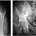
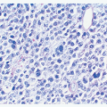
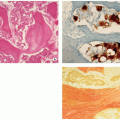
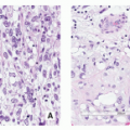

Stay updated, free articles. Join our Telegram channel

Full access? Get Clinical Tree


