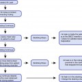
Metallic taste
Tinnitus
Light-headedness
Anxiety
Somnolence
Seizure
Coma
Cardiovascular collapse
Death
Table 6.2
Maximum allowable doses of local anesthetics
LA | Effects | |
|---|---|---|
Cocaine | 1.0 mg/kg | Hypertension, tachycardia, MI |
Lidocaine | 5.0–7.0 mg/kg | Seizure, ventricular arrythmia, coma |
Benzocaine | 1–2 sprays | Methemoglobinemia |
Several local anesthetics have specific side effects. Cocaine, for example, acts as a sympathomimetic and can lead to hypertension, tachycardia, coronary vasospasm, and myocardial infarction. Benzocaine, which is metabolized in the blood, can lead to the formation of methemoglobin, which is toxic at levels above 70%. Methemoglobinemia can result in cyanosis, arterial desaturation, and death. As little as a single spray of benzocaine has resulted in toxic levels of methemoglobin. The treatment for methemoglobinemia involves supplemental oxygenation and the use of intravenous methylene blue. Thus, cocaine and benzocaine are associated with serious side effects – side effects that require judicious use and deserve cautious practice. In our department, we almost never find cocaine necessary and infrequently rely on benzocaine.
While the side effect profile as well as the substance abuse potential for cocaine may preclude its regular use as a local anesthetic, benzocaine remains valuable in every day bronchoscopic practice, due to its rapidity of action. The vasoconstrictive advantages of cocaine can be achieved with such agents as phenylephrine nasal spray, making cocaine less essential. Therefore, the therapeutic use of cocaine in interventional pulmonary procedures may have days that are numbered.
Overview of the Range of Sedation
Moderate and deep sedation represent the middle range of a spectrum of anesthetic depths that include light sedation (or anxiolysis), moderate sedation (or conscious sedation), deep sedation and general anesthesia (see Table 6.3). The differences in the sedation depth are evident by the difference in the way that patients respond to stimuli. Under moderate sedation, for example, patients demonstrate preserved protective airway reflexes. Also under moderate sedation, patients show appropriate responses to verbal or light tactile stimulation. Under deep sedation on the other hand, the patient may lose the protective airway reflexes and may not respond to verbal or light tactile stimuli. Patients under deep sedation may respond to repeated or painful stimuli. The choice to use either moderate or deep sedation techniques depends on the procedure planned, the experience of the pulmonologist, the experience of the sedation nurse or anesthetist, and the patient’s comorbidities and concerns.
Table 6.3
The range of depths of anesthesia
Analgesia | Minimal | Moderate | Deep | GA |
|---|---|---|---|---|
Minimal sedation | Moderate sedation | Deep sedation | General anesthesia | |
Preserved airway reflexes | Preserved airway reflexes | Protective reflexes may not be preserved | Reflexes not preserved | |
Appropriate response to verbal stimulus | Appropriate response to verbal or tactile stimulus | Unable to respond to verbal or light tactile stimulus | ||
May respond to repeated or painful stimulus | No purposeful responses to painful stimulus | |||
Spontaneous ventilation | Spontaneous ventilation | Ventilation may be inadequate | Ventilation may be inadequate | |
MAC | GA | |||
Studies that look at whether sedation techniques can improve bronchoscopic procedures are conflicting. Some studies show that moderate sedation can improve the patient outcomes for bronchoscopic procedures. Other studies show no improvement. One study by Putinati et al. identified as much as half the serious complications as coming from unwanted effects of the sedation. From a review of the literature, one can conclude that there is no conclusive evidence to argue for the administration of one sedation technique over another. The final decision must include the consideration of the safety and comfort of the patient, the experience of the pulmonologist and the anesthetist, and the anatomic requirements of the procedure.
Requirements to Provide Sedation
In the United States, the Joint Commission requires that all clinicians who administer sedation for interventional pulmonary procedures adhere to nine standards:
1.
A presedation patient assessment is performed.
2.
The sedation risks and options are discussed.
3.
Moderate or deep sedation is provided by qualified personnel.
4.
Moderate or deep sedation planes are written as a set of sedation orders.
5.
Each patient’s physiologic status is monitored during the sedation.
6.
Each patient’s physiologic status is collected and analyzed.
7.
Qualified personnel are used to provide moderate or deep sedation.
8.
Each patient’s postprocedure status is assessed on admission to and before discharge from the postsedation recovery area.
9.
Qualified personnel are used to discharge patients from the postsedation recovery area, and this should be according to criteria approved by the medical staff.
The presedation assessment requires a standard history and physical exam with certain information that directly relates to the safe conduct of moderate or deep sedation. This information includes:
1.
Assessment of the airway.
2.
Evaluation of comorbidities that could negatively affect the safety of the sedation plan. Any prior reaction to sedation will be noted.
3.
Determination of the ASA physical status score.
4.
Determination that the patient has complied with NPO guidelines. These guidelines are often institution-specific. We use NPO from solids for 6 h and NPO from clear liquids for 2 h prior to the procedure, unless the patient has a reason to have a full stomach (trauma, reflux, hiatal hernia).
5.
Determination that the patient has a reliable escort home (if outpatient).
6.
Consent for sedation.
The assessment of the airway is crucial because it helps to identify those patients in whom maneuvers to establish airway patency and to perform intubation may be difficult. These maneuvers can become necessary if the patient becomes oversedated. The Mallampati classification is one method to categorize patients who may prove difficult to intubate. To perform this examination, have the patient open his or her mouth fully and extend the tongue without phonating. The extent to which the palate and tonsils are visible determines the Mallampati classification (Fig. 6.1):
Class I: The entire tonsillar pillars are visible.
Class II: The top half of the tonsillar pillars is seen.
Class III: The base of the uvula is seen.
Class IV: The hard palate is seen.
Fig. 6.1
Mallampati classification
In addition to the Mallampati classification, there are several other anatomic predictors of difficult intubation and by inference, difficulty in rescue from oversedation. A receding mandible, limited mouth opening of less than 3.5 cm between the upper and lower incisors, pronounced overbite and/or an inability to prognath the jaw, a decreased thyromental or hyomental distance, a history of obstructive sleep apnea, and a history of a difficult or failed intubation. Patients who possess one or more of these anatomic predictors of a difficult intubation may benefit by having their sedation provided by an anesthesiologist.
Comorbid conditions can also influence the safety and conduct of moderate or deep sedation. A general assessment of the impact of comorbidities on operative mortality is the ASA physical status. Based on the patient’s comorbid conditions and their impact on the patient’s daily living, the ASA physical status consists of the following categories:
Class I: No disturbances
Class II: Mild to moderate systemic disturbances
Class III: Severe systemic disturbance that impacts daily function
Class IV: Life-threatening systemic disturbance
Class V: Moribund, not expected to survive
While a general assessment is important, consideration of the risks and potential complications resulting from specific comorbidities must also be considered. Conditions that limit the patient’s cognitive function such as stroke and dementia can make the patient more sensitive to sedative medications. Patients with severe COPD more often experience the side effects of respiratory depression and respiratory failure due to opioids and benzodiazepines, for example. Cardiac problems such as uncompensated congestive heart failure and aortic stenosis can result in hemodynamic instability following modest doses of sedative/hypnotics. Liver and kidney disease can affect the metabolism and excretion of many drugs and can result in exaggerated and prolonged effects. While a detailed discussion of the specific effects of comorbidities is beyond the scope of this chapter, the pulmonologist should respect the potential problems that such comorbidities can present.
Equipment and Monitors
The safe provision of moderate or deep sedation requires the equipment necessary to provide the next deeper level of sedation, namely, general anesthesia. Such rescue equipment is listed in Table 6.4. The availability, functionality, and sterility (where applicable) of the equipment should be assessed regularly, and the providers of sedation should be familiar with the location of each item.
Table 6.4
Equipment for moderate/deep sedation
Category | Specification |
|---|---|
Intravenous access | IV fluids |
IV tubing, cannulae | |
IV catheters | |
Gloves | |
Tourniquets | |
Alcohol wipes | |
Tape | |
Monitoring | Noninvasive blood pressure device |
Pulse oximeter | |
Capnograph | |
Electrocardiograph | |
Airway | Oxygen source, tubing, cannulae |
Bag/mask | |
Suction | |
Oral and nasal airways | |
Laryngoscopes | |
Endotracheal tubes | |
Laryngeal mask airways | |
Emergency | Reversal agents (naloxone, flumazenil) |
ACLS medications | |
Cardiac defibrillator |
Although essential equipment is important, the most crucial element to providing safe sedation is a vigilant and trained sedation provider. This provider, if a nurse, should have no other significant clinical duties during the procedure and should have the same level of training and privileges in providing sedation as the physician performing the interventional pulmonology procedure. Inadequate monitoring of patients has been a frequent cause of adverse events during bronchoscopy. Vital signs (heart rate, respiratory rate, blood pressure, oxygen saturation) should be recorded every 5 min along with level of consciousness and skin condition. Temperature should be documented at regular intervals. In addition, the timing and doses of individual drugs should be recorded along with the indications and effectiveness. The administration of intravenous fluids should be quantified. While the respiratory rate can be obtained through the measure of chest impedance using electrocardiogram electrodes, this author prefers the use of carbon dioxide monitors as a method to not only obtain respiratory rate but also to document the continuous production of CO2 as well as the continuous patency of the airway (See Fig. 6.2).

Fig. 6.2
Normal capnogram
Related posts:


Stay updated, free articles. Join our Telegram channel

Full access? Get Clinical Tree


