Advances in MR imaging technologies, as well as the widening of their availability, boosted their use in the diagnosis of spinal disorders and in the preoperative planning of spine surgeries. However, the most consolidated approach to the assessment of adult patients with spinal disorders is based on the analysis of full standing radiographs (posteroanterior and laterolateral views). In this article, the radiographic spinal and pelvic parameters, which have relevance in the clinical management of adults with spinal disorders, are summarized.
Key points
- •
The pelvis is the keystone of spinal alignment.
- •
Full standing radiographs (posteroanterior and laterolateral views) are necessary for a proper analysis of the spinopelvic alignment.
- •
The use of MR imaging alone may not be sufficient for correct pre-operative planning, but should be supplemented by the analysis of the global spinal alignment.
Discussion of problem
Advances in MR imaging technologies as well as the widening of their availability boosted their use in the diagnosis of spinal disorders and in the preoperative planning of spine surgeries. As a matter of fact, MR imaging provides excellent contrast between different types of soft tissues as well as a qualitative estimate of the tissue composition, for example, the water and fibrous contents of the intervertebral disc. These qualities undoubtedly allow for improvements in the diagnosis of various degenerative disorders and facilitate the selection of the optimal treatment. Therefore, MR imaging is often considered the imaging modality of choice in the management of many degenerative disorders of the spine, especially if the symptoms seem to be caused by a single or a couple of motion segments.
Besides, the analysis of planar radiographs of adults with spinal disorders has a long and consolidated tradition in the orthopedic community. Even if taking into account the limited capability of standard radiographs in providing clinically valuable information about soft tissues, the excellent imaging quality of bone tissue together with the possibility of assessing the spinal alignment under the action of a physiologic load (typically the standing posture) provides clear advantages with respect to MR imaging, which is inherently limited by a lower resolution and the horizontal posture of the patient. Thus, it is thought that a combined use of the 2 imaging modalities may be advantageous in cases in which both the clinical status of the soft tissues and the spinal alignment must be taken into account in order to conduct an optimal preoperative planning.
In this article, the radiographic spinopelvic parameters that have relevance in the clinical management of adults with spinal disorders are summarized. One case, in which a preoperative planning based only on MR images would have possibly led to a suboptimal surgical solution, is then presented and discussed in depth.
Anatomy
Spinopelvic Alignment
In 1992, a radiological article, which had a decisive impact on spine surgery in the 2 following decades, was published. This work introduced for the first time the relationship between the pelvis (through anatomic and functional pelvic parameters) and the sagittal alignment of the spine, and furthermore, the tendency to maintain the body in the most economical position in terms of energy consumption and vertebral strain. The 3 main pelvic parameters that were identified were the pelvic incidence (PI), the pelvic tilt (PT), and the sacral slope (SS) ( Fig. 1 ).
PI is the only one of the 3 parameters that has a fixed anatomic value for each individual and is therefore not dependent on the posture; indeed, it is usually referred to as an anatomic parameter. Several studies assessed the distribution of PI in the general population and reported a marked variability. PI approximately ranged between 35° and 85°, with a tendency toward an increase during growth. In these studies, patients with lower PI tended to have a flatter profile of the back, especially a lower curvature of the lumbar spine. The wide distribution of PI in the population did not allow for the determination of clear “healthy” and “pathologic” categories, because the spine curvature seemed to be able to adapt efficiently to the various anatomic morphologies of the pelvis.
PT and SS do not have fixed values in a specific patient, but are dependent on the posture. Indeed, a flexion of both hip joints would lead to a decrease of PT and an increase of SS, whereas PI would be not affected. Both parameters tend to change during the life of the subject, especially during childhood.
Other important descriptors of the spinal shape are the regional curvatures, that is, cervical lordosis (CL), thoracic kyphosis (TK), and lumbar lordosis (LL) ( Fig. 2 ). The physiologic curvature of the cervical spine is still a matter of debate, because studies reported an absence of lordosis or a small kyphosis in asymptomatic subjects, whereas well-known studies reported a physiologic lordotic range of 15° to 20°. Similarly, TK was shown to have a wide range in asymptomatic subjects, and an average value in the general population of 40°. As mentioned above, the value of LL is strictly dependent on the pelvic morphology, with subjects having high PI showing correspondingly high LL. There is general agreement about the fact that there is a strong interplay between the regional curvatures of the spine as well as with the pelvis morphology, and a correct analysis of each of these parameters cannot disregard the other regional curvatures.
In order to characterize the global sagittal spinal alignment, other radiological parameters have been introduced and are currently widely used (see Fig. 2 ). The sagittal vertical axis (SVA), also named C7 plumb line, describes the distance in the horizontal direction between a vertical line through the vertebral body of C7 and the posterosuperior corner of the sacral plate. SVA is commonly used to describe with a single measurement the global sagittal alignment of the thoracolumbar spine. Values less than 5 cm have been reported as physiologic, whereas higher SVAs characterize sagittal malalignment. It should however be noted that SVA is not a strictly anatomic parameter, but depends on posture.
The difference between PI and LL (PI − LL) is also often used as a single descriptor of the spinal alignment. As LL should adapt to the pelvic morphology (described with PI), an excessive mismatch between the 2 values would represent a condition in which the patient is not able to find a spinopelvic organization fitting well her or his pelvic anatomy. It was suggested that a value of PI − LL lower than 10° would indicate a problematic alignment, although the same investigators indicated that this rule should not be interpreted strictly. These observations are commonly used as a base to determine the target correction in the surgical treatment of sagittal malalignment.
Another radiological parameter that emerged recently to analyze the spinal alignment in the sagittal plane is the pelvic shift (P. Sh.) (see Fig. 2 ), which describes the posterior offset of the pelvis with respect to the heels. This parameter is especially useful to characterize the compensatory mechanisms adopted by patients with alignment disorders in order to regain a sustainable balancing.
Sagittal Imbalance and Compensatory Mechanisms
When pathologic changes to the alignment of single segments or longer regions occur, the human body may react with a compensatory mechanism to counterbalance it, or with a combination of different mechanisms. Typical degenerative changes that may induce compensation are the loss of LL (even on 1 or 2 levels due to degenerative disc disease), and the increase of kyphosis in the thoracic spine. It is thought that the aim of compensatory mechanisms is to reduce the energy expenditure in standing and during daily activities. Indeed, an imbalanced posture requires a significant constant activation of the trunk muscles in order to counterbalance the weight of the upper body, which generates a moment with respect the feet due to its forward inclination.
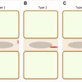
One of the first compensatory mechanisms that frequently occur also in asymptomatic elderly subjects is the retroversion of the pelvis, which corresponds to a decrease of SS and an increase of PT ( Fig. 3 ). In addition, single lumbar segments may exhibit hyperextension or retrolisthesis, thus creating a risk of stenosis of the spinal canal. Another compensatory strategy detectable, especially in younger subjects having a good spine flexibility, is the reduction of TK, which combined with the loss of LL results in a relatively flat sagittal profile of the spine. More severe cases show a flexion of the knees, which induces additional energy expenditure due to the recruitment of the thigh muscles and may generate anomalies in the gait pattern.