The human spine is a complex biomechanical system composed of multiple articular structures controlled by muscles. Spine diseases are frequently related to a loss of stability. Dedicated imaging protocols have been developed to evaluate spinal instability. Dynamic radiography with lumbar flexion-extension is used most often; however, in traumatic instability, computerized tomography provides better diagnostic accuracy for fracture detection. Novel technology improvements allow acquisition of dynamic MRI with axial load or upright standing techniques to simulate a more pathologic condition compared with conventional supine scans. This article reviews the basic concepts of spinal instability and describes the role of different imaging techniques in its assessment.
Key points
- •
Degenerative, traumatic, and neoplastic instabilities are based on different pathophysiologic mechanisms, so each pattern requires a peculiar integrated clinical-radiologic approach.
- •
Dynamic radiographs with upright true lateral neutral-flexion-extension projections are still the most widely used imaging approach to diagnose instability in the daily practice.
- •
In traumatic instability, computerized tomography (CT) is the preferred image modality because it is able to detect quickly even the tiniest unstable fractures with reduced patient manipulation.
- •
Conventional MRI acquired in supine rest position often correlates poorly with clinical findings because of the loaded positional dependence of patient symptoms.
- •
Novel dynamic MRI approaches simulate closely the pathologic conditions that elicit symptoms in patients with instability, providing strict linkage between clinical status and imaging.
Introduction
The human spine is a complex biomechanical system composed of multiple articular structures controlled by muscles. It has 2 principal crucial functions: support and protection. The spine supports the head and trunk and protects the spinal cord, nerve roots, and vertebral arteries during every movement. Furthermore, it transfers power forces between upper and lower limbs.
These functions presuppose spine stability; however, even though several articles have been published about this concept, a consensus definition of stability is still lacking.
Clinical problems of the human spine continue to be prevalent in society. Examples include low back pain, sciatica, spinal deformity in both adults and children, spinal tumors, and spinal injury, including trauma to the spinal cord. Frequently these are related with a loss of stability (instability) particularly at the lumbar level. Traumatic, neoplastic, and degenerative instability are important causes of spinal pain and disability.
Conventional radiology with dynamic projections has long been considered the technical reference to assess the degree of instability, and different methods have been developed to assess the presence of listhesis; however, radiography has proved inadequate to assess stability in case of spinal fractures. Furthermore, because patients with instability frequently present concomitant disk and radicular pathologies, conventional radiography is limited by its diagnostic use in the assessment of these structures. CT, instead, yields high-resolution reconstructions in every spatial plane to detect even the tiniest fractures, revealing potentially unstable lesions.
MRI is the only imaging modality that directly assesses ligaments integrity, which is crucial for spine stability. In the past 2 decades, novel loaded MRI systems have been developed to evaluate instability in a more realistic pathologic condition, adding relevant information to conventional supine MRI.
This article reviews the basic pathologic concepts of spinal instability and describes the role of the different imaging techniques in the assessment of this clinical issue.
Pathologic principles of instability
Spinal stability depends on the interaction of 2 strictly related elements, column and muscles, under the control of the central nervous system. The loss of stability leads to instability, because for stability, a univocal definition has not been agreed on, even if it remains critical in the surgical decision-making process. White and Panjabi defined instability as “the loss of the ability of the spine under physiologic loads to maintain its patterns of displacement so there is no initial or additional neurologic deficit, no major deformity, and no incapacitating pain.” Pope and colleagues intended instability as a loss of stiffness leading to abnormal and increased movement in the motion segments. Spinal movements are 3-D with coupled movements, so spine instability always causes dysfunctional motions in more than 1 direction.
There are different patterns of instability based on the pathophysiologic mechanisms that sustain the process: degenerative, traumatic, and neoplastic.
Degenerative Instability
Spinal degenerative instability is a common cause of pain and disability. This pathologic process starts with the lesion of a component of the column, leading to an inappropriate response of the muscles and consequently an erroneous positional feedback of the column. In this way, a vicious circle causes a chronic dysfunction and pain through 3 steps, the degenerative cascade: dysfunction, instability, and restabilization.
Dysfunction phase
The dysfunction phase is characterized by an occasional undefined low back pain, with no or minimal changes in the spinal joints; frequently in this situation no imaging findings are appreciable.
Instability phase
In the instability phase, back pain becomes more and more frequent to chronic. Multiple signs are appreciable on radiologic examinations (x-ray, MRI, and CT scans), such as facets degeneration and disk space narrowing. These elements lead to abnormal vertebral movement and alignment, up to anterolisthesis or retrolisthesis. At the beginning of the instability phase, the process is usually limited to a single joint but then, it involves the adjacent joints, resulting in a multifocal pathology. End plate, peduncle, and isthmic edema; Modic changes; traction spurs; extended discal vacuum; facets gapping with joint effusion or vacuum; synovial cysts; annular tears; spondylolysthesis; and retrolysthesis are typical imaging findings of the full-blown disease. Sometimes on standard supine scans, signs of instability are available but vertebral alignment is preserved; however, this alignment is a misleading condition due to the absence of load bearing. In these cases, a dynamic radiograph or an upright MRI is suitable to diagnose occult instability.
Restabilization phase
In the final phase, restabilization, structural compensatory remodeling phenomena bring reduced mobility and stiffness. Marginal osteophytes; disk collapse; radial expansion of vertebral bodies and facets; and end plate, spinous, and transverse sclerosis: all these remodeling processes interrupt vertebral slippage but also block physiologic movements.
Traumatic Instability
Unlike degenerative instability, the relationship between imaging findings and clinical symptoms tends to be more direct in traumatic spinal instability. Every time a trauma damages a column element, it produces a certain degree of instability; all spinal components contribute to stability. Different studies have analyzed the effects of trauma on the spine and different models have been developed. Denis proposed a model formed by 3 vertical columns: an anterior column, including the anterior halves of the bodies and disks with the adjacent anterior longitudinal ligament; a middle column, including the posterior half of the bodies and disks with posterior longitudinal ligament; and a posterior column, consisting of neural arches and the posterior ligamentous complex, including the supraspinous, interspinous, and flava ligaments and facet joint capsules. Denis assessed that instability was due to the simultaneous failure of at least 2 columns, creating situations of instability. Today Denis’s model remains among the most accepted references.
Although a considerable amount of energy is required to produce the first injury in a vertebra, just a small additional trauma is sufficient to convert a lesion from stable to unstable and to switch from conservative treatment to surgical stabilization.
The thoracolumbar spine is the most common site afflicted by trauma; L1 is the most common vertebra followed by T12. Its predilection and vulnerability for injury stems from the following reasons: (1) mobility: the thoracolumbar spine presents a high degree of mobility, so is subjected to all kinds of forces, resulting in a district highly vulnerable to fractures; (2) transition zone: the thoracolumbar region is relatively straight (kyphosis from 0°–10°) and situated between the kyphotic thoracic and lordotic lumber spine; unlike the thoracic spine, the absence of costovertebral structures no longer protects the thoracolumbar zone; and (3) facet joints: facet joints of the thoracic spine are coronally oriented to resist flexion-extension and that of lumbar spine are sagittally oriented to allow flexion-extension. In the thoracolumbar area, facet joints show a transition from predominantly coronal to predominantly sagittal orientation.
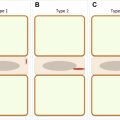
Thoracolumbar fractures have been classified by Magerl and colleagues into 3 main groups: A, B, and C types (each 1 divided into 3 subgroups), corresponding to increasing degrees of instability; this classification system is based on progressive morphologic damage determined by 3 fundamental forces: compression, distraction, and axial torque (rotation). Type A injuries are either stable or partially compromised but never completely unstable, because the posterior longitudinal ligament is intact, while the degree of stability progresses from the most stable type A to the most unstable type A3; types B1 and B2 are unstable in flexion because of the posterior ligament injury while stability in the extension is maintained by an intact anterior ligament; type B3 injuries are unstable in extension but may be stable in flexion, if the posterior ligament is intact; and all type C injuries are unstable.
The most important finding in recognizing stable versus unstable fractures is the state of the posterior ligaments; the status of posterior ligaments after an injury is of great importance for the stability of the injured spine. In burst fractures, the condition of the posterior column suggests fracture instability, which increases remarkably in cases of lesions to the posterior ligaments.
MRI is the only imaging modality able to assess changes in ligaments with significant accuracy and has a crucial role in treatment choice. MRI interpretation in this clinical scenario, however, needs to be improved. It does not find clear signs of ligament discontinuity but just signal modifications occurring in and around apparently continuous ligaments: the clinical meaning of these findings is still unclear. On the other hand, a ligament must not necessarily be torn to become biomechanically ineffective, and the absence of signal modifications cannot always exclude instability.
Neoplastic Instability
Spinal instability as a result of a neoplastic process differs significantly from high-energy traumatic injuries in the pattern of bony and ligamentous involvement, potential for healing, neurologic manifestations, and bone quality. It requires a specific and different set of criteria for stability assessment.
Neoplastic spine instability has been defined by the Spine Oncology Study Group as loss of spinal integrity as a result of a neoplastic process associated with movement-related pain, symptomatic or progressive deformity, and/or neural compromise under physiologic loads.
Spinal Instability Neoplastic Score
A panel of expert members from Spine Oncology Study Group developed the Spinal Instability Neoplastic Score, taking into account 6 components, each receiving a score that rises based on the rate of instability. The minimum score is 0 and maximum is 18. Scores of 0 to 6 denote stability, scores of 7 to 12 denote indeterminate (possibly impending) instability, and scores of 13 to 18 denote instability. Patients with Spinal Instability Neoplastic Score scores of 7 to 18 warrant surgical consultation. The 6 components are spine location, mechanical pain, bone lesion quality, spinal alignment, vertebral body collapse, and posterolateral involvement of spinal elements.
Impending spinal instability affects clinical decision making in oncologic spinal disease. In patients with neoplastic spinal disease without neurologic deficit, it is crucial to recognize which situations are unstable or may lead to spinal instability and neurologic injury. This allows proper stabilization of patients with severe mechanical pain and hopefully prevents painful collapse with neurologic consequences.
Imaging in spinal instability
Various techniques have been developed over the past decades to evaluate the presence of spinal instability both clinically and radiologically. The most widely used approach is lumbar flexion-extension radiography but CT and MRI are also applied in this field with dedicated protocol.
Dynamic Radiography
Radiographs are acquired in dynamic modality with upright anterior-posterior and true lateral neutral-flexion-extension projections ( Fig. 1 ).
Related posts:


Stay updated, free articles. Join our Telegram channel

Full access? Get Clinical Tree


