Suresh K. Mukherji, MD, FACR, Consulting Editor
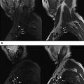
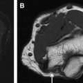
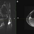
This unique and comprehensive edition contains state-of-the-art articles that describe nerve anatomy, pathophysiology, approach to MRN imaging, and interpretation. This issue is “image-rich” with numerous examples of pathology involving the brachial plexus, lower extremity, and lumbosacral plexus. Other articles focus on MR-guided perineural interventions, peripheral nerve surgery approaches, and postoperative imaging.
I wish to thank Dr Chhabra for accepting this challenging topic and all of the contributors for their outstanding contributions. This is truly a unique edition that will be a valuable resource for many years to come!
Stay updated, free articles. Join our Telegram channel

Full access? Get Clinical Tree


