
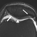
Due to its high spatial resolution, multiplanar capability, and excellent tissue contrast, MRI is the noninvasive modality of choice for evaluating articular cartilage in both the clinical and the research setting. Cartilage imaging in the past has focused primarily on large joints such as the knee and hip. However, with recent development of high field strength scanners, three-dimensional cartilage-sensitive sequences, and multichannel coils, images of joints with higher spatial resolution, thinner slices, and greater tissue contrast can be obtained in clinically feasible scan times. With this new technology, MRI is now being used to evaluate the articular cartilage of the shoulder, elbow, ankle, and small joints of the upper and lower extremity.
This edition of Magnetic Resonance Imaging Clinics of North America gives a broad overview of the role of MRI in the evaluation of articular cartilage. Articles in this issue describe the role of histological structure on the MRI appearance of normal articular cartilage and discuss currently used MRI techniques to evaluate both cartilage morphology and cartilage physiology. Factors related to rapid progression of osteoarthritis and the use of MRI in evaluating patients in osteoarthritis research studies and following cartilage repair procedures are also revewed. Articles discussing the role of MRI in the evaluation of patients with inflammatory, crystalline-induced, and infectious arthritis and patients with various articular disorders of the hip, knee and ankle, and upper extremity are included.
I express my utmost appreciation to the authors who have spent much time and effort writing these articles. I was fortunate to receive the participation of many excellent musculoskeletal radiologists who are international experts in the area of cartilage imaging. I have enjoyed reading these articles and have learned a great deal about cartilage imaging in my role as guest editor. I have no doubt that the readers will do so as well.
Related posts:
 Morphologic Imaging of Articular Cartilage
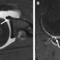
 MR Imaging Assessment of Articular Cartilage Repair Procedures
 Magnetic Resonance Imaging in Knee Osteoarthritis Research: Semiquantitative and Compositional Assessment
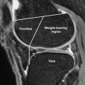
MR Imaging of the Articular Cartilage of the Knee and Ankle
MR Imaging of Early Hip Joint Degeneration
Magnetic Resonance Imaging in Knee Osteoarthritis Research: Semiquantitative and Compositional Assessment
MR Imaging of the Articular Cartilage of the Knee and Ankle
MR Imaging of Early Hip Joint Degeneration
 The Current State of Imaging the Articular Cartilage of the Upper Extremity
Stay updated, free articles. Join our Telegram channel

Full access? Get Clinical Tree


