Table 15-1.
Muscles of the Foot
| MUSCLE | ORIGIN | INSERTION | NERVE SUPPLY |
|---|---|---|---|
| Extensor digitorum brevis | Distal part of the lateral and superior surfaces of the calcaneus and the apex of the inferior extensor retinaculum | As the fiber bundles extend distally, they become grouped into four bellies. Those fibers of the most medial and largest belly are known as extensor hallucis brevis. The tendon of this muscle inserts onto the base of the first metatarsal. The remaining fiber bundles have more variable insertions. The second toe fibers usually insert onto the middle dorsal aspect of the base of the proximal phalanx and are often united with the long extensor tendon. The three more lateral tendons are usually fused with the lateral margins of the corresponding long extensor tendons near the bases of the respective middle phalanges and usually extend to the bases of the corresponding proximal phalanges. | Deep peroneal |
| Flexor digitorum | Medial process of the tuber calcanei, posterior third of the plantar aponeurosis, and medial and lateral intermuscular septa | Tendons of the short (brevis) flexors pass superficial to those of the long flexor into the osteofibrous canals on the flexor surface of the digits. On the proximal phalanx of each toe, the tendon of the short flexor divides and forms an opening through which the tendon of the long flexor passes. The tendons of the short flexor insert onto the base of the middle phalanx | Surface near the medial edge of the muscle |
| Quadratus plantae (flexor accessorius) | Two heads: a small lateral and a large medial one. (1) Lateral head arises from an elongated tendon from the lateral process of the tuberosity of the calcaneus and from the lateral margin of the long plantar ligament. (2) Medial head originates from the medial surface of the calcaneus in front of the tuberosity and from adjacent ligaments | The two heads are separated at their origin by a short triangular space. The heads fuse to form a single belly, but the fiber bundles of each head are separately inserted. From the lateral head, the fibers insert into the lateral margin of the flexor tendon. The medial head inserts as an aponeurosis into the deep surface of the flexor tendon | Lateral plantar nerve branch that passes obliquely across the superficial surface of the muscle, parallel with the tendon of the flexor digitorum longus |
| Lumbricals | Three lateral lumbricals arise from the adjacent sides of the digital tendons of the flexor digitorum longus. The first lumbrical arises on the medial margin of the second toe | Fiber bundles of each muscle converge on both sides of a tendon that becomes free near the metatarsophalangeal joint and is inserted onto the medial side of the proximal phalanx of the appropriate toe. A tendinous expansion is inserted into the aponeurosis of the extensor muscle | Three lateral lumbricals are usually supplied by branches of the deep ramus of the lateral plantar nerve. Medial lumbrical is supplied by the first common plantar digital branch of the medial plantar nerve. This nerve may supply the two more medial muscles, or the medial muscles may receive a double nerve supply |
| Abductor hallucis | Medial border of the medial process of the tuber calcanei, flexor retinaculum, and plantar aponeurosis | Along with the tendon of the medial belly of flexor brevis onto the base of the proximal phalanx of the great toe | Branch of the medial plantar |
| Flexor hallucis brevis | From the plantar surface of the lateral cuneiform and cuboid bones | The medial and lateral sides of the base of the proximal phalanx of the great toe | Branch from the medial plantar or first plantar digital. Rarely, the lateral body may receive a branch from the lateral plantar |
| Adductor hallucis, oblique head | Tuberosity of the cuboid and sheath of the tendon of the peroneus longus, the plantar calcaneocuboid ligament, the third cuneiform, and bases of the second and third metatarsals | By a flat tendon that inserts in common with that of the flexor brevis onto the lateral part of the plantar surface of the base of the proximal phalanx, and by a slip into the aponeurosis of the long extensor muscle on the back of the great toe | Branch of the deep ramus of the lateral plantar |
| Adductor hallucis, transverse head | Joint capsules of the third, fourth, and fifth metatarsophalangeal joints and the deep transverse metatarsal ligaments | By a common tendon that splits and passes on each side of the tendon of the oblique head and is inserted into the sheath on the tendon of the long flexor of the great toe | Branch from the deep ramus of the lateral plantar |
| Abductor digiti minimi | Lateral and medial processes of the tuber calcanei and lateral and plantar surfaces of the body of the bone in front of these, lateral intermuscular septum, deep surface of the lateral plantar fascia, and fibrous band extending from the calcaneus to the lateral side of the base of the fifth metatarsal | Onto the lateral surface of the proximal phalanx of the little toe and the metatarsophalangeal capsule | Lateral plantar |
| Flexor digiti minimi brevis | Sheath of the peroneus longus, tuberosity of the cuboid, and base of the fifth metatarsal | By short tendinous bands onto the base of the proximal phalanx of the little toe, the capsule of the corresponding joint, and the aponeurosis on the dorsal surface of the toe | Branch from the superficial ramus of the lateral plantar |
| Opponens digiti minimi | An inconstant muscle, it may arise from the sheath of the peroneus longus and the tuberosity of the cuboid by a thin tendon that passes over the tuberosity of the fifth metatarsal | Onto the lateral surface of the fifth metatarsal | Branch from the nerve to the flexor brevis and the superficial ramus of the lateral plantar |
| Interosseous, dorsal | Each of the three lateral dorsal interosseous muscles arises from the sides of the shaft and the plantar surface of the bases of the metatarsals, bounding the space in which each lies, from the fascia covering it dorsally, and from the fibrous prolongations from the long plantar ligament. The first (medial) has a similar origin, except that its medial origin is by a tendinous slip from the peroneus longus tendon and, occasionally, by fiber bundles from the medial side of the proximal end of the first metatarsal | The first and second interosseous muscles onto each side of the base of the proximal phalanx of the second toe; the third and fourth onto the lateral side of the bases of the proximal phalanges of the third and fourth toes. Each tendon adheres to the capsule of the adjacent joint | Deep branch of the lateral plantar. The interosseous muscles of the fourth interspace are usually supplied by a branch from the superficial ramus of the lateral plantar |
| Interosseous, plantar | The plantar interosseous muscle arises from the proximal third of the medial plantar surface of the shaft, from the base of the metatarsal on which it lies, and from the fascial expansions of the long plantar ligament | Onto a tubercle on the medial side of the base of the proximal phalanx of the digit to which it goes | Lateral plantar |
Axial
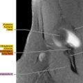
Figure 15.1.1

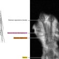
Figure 15.1.2