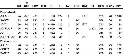
BRAIN WITHOUT AND WITH CONTRAST
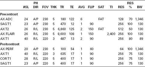
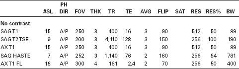
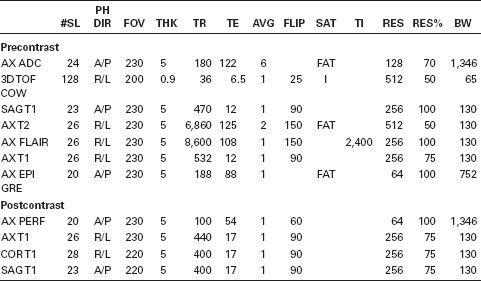
#SL, number of slices; PH DIR, phase direction; FOV, field of view in millimeters; THK, slice thickness in millimeters; TR, repetition time in milliseconds; TE, echo time in milliseconds; AVG, number of averages; FLIP, flip angle; SAT, type of saturation; TI, inversion time in milliseconds; RES, resolution in phase-encoding direction; RES%, resolution in frequency-encoding direction expressed as percentage of resolution (100% results in rectangular FOV); BW, bandwidth.


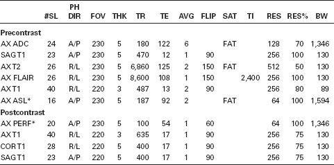
*ASL, arterial spin labelled perfusion may be used additionally to contrast enhanced perfusion or as a substitute to it, *see specific perfusion protocols.

*ASL, arterial spin labelled perfusion, *see specific perfusion protocols.
Related posts:
Stay updated, free articles. Join our Telegram channel

Full access? Get Clinical Tree


