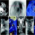
Fig. 64.1
CT-PET shows partial resection the lower left lung lobe, with centimetric contextual fibrotic tissue thickening and striae—scars that reach the hilum; mild carbohydrate consumption of the lesion
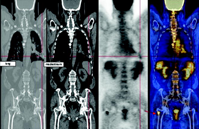
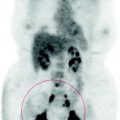
Fig. 64.2
The coronal reconstructions show a bone-producing lesion to the greater trochanter of the right femur, that has limited metabolism
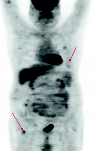
Fig. 64.3
 Laryngeal Squamous Carcinoma: Staging
 Radio-Treated Cancer of the Posterior Hemi-Circumference of the Anal Canal: Post-Actinic Fibrosis
 Bone-Destroying Metastases in Thyroid Undifferentiated Carcinoma
 Surgically Treated Endometrial Cancer: Bilateral Nodal Recurrence
 Undifferentiated Gastric Adenocarcinoma with Peritoneal Carcinosis
 Bone Metastases from Breast Cancer: Progression of Disease and Subsequent Response to Radiotherapy


MIP image in patients with liver transplantation, FDG-PET examination frequently shows nonspecific background high uptake due to the slow clearance of the tracer, especially in the abdomen. As immunosuppressed patients have a higher incidence of new cancers and are therefore considered a high-risk category, some authors suggest surveillance with CT-PET
Related posts:
Laryngeal Squamous Carcinoma: Staging
Radio-Treated Cancer of the Posterior Hemi-Circumference of the Anal Canal: Post-Actinic Fibrosis
Bone-Destroying Metastases in Thyroid Undifferentiated Carcinoma
Surgically Treated Endometrial Cancer: Bilateral Nodal Recurrence
Undifferentiated Gastric Adenocarcinoma with Peritoneal Carcinosis
Bone Metastases from Breast Cancer: Progression of Disease and Subsequent Response to Radiotherapy
Stay updated, free articles. Join our Telegram channel
Full access? Get Clinical Tree
