Mucocele of the Appendix
Michael P. Federle, MD, FACR
Key Facts
Terminology
Chronic cystic dilatation of appendiceal lumen by mucin accumulation
Imaging
Classified into 3 groups based on histology
Mucosal hyperplasia (simple mucocele)
Mucinous cystadenoma (most common)
Mucinous cystadenocarcinoma (↑ risk of perforation)
Pseudomyxoma peritonei
Due to rupture: Malignant > benign mucocele
Peritoneal cavity filled with mucus seedlings
Loculated ascites; scalloped surface of liver and spleen
Myxoglobulosis
Rare variant with multiple small globules ± calcifications
Mucocele
Calcification (curvilinear) within wall or lumen
Mucinous cystadenocarcinoma
Large irregular mass with thickened nodular wall
Top Differential Diagnoses
Acute appendicitis (abscess)
Appendiceal tumors
Cecal carcinoma
Ovarian cystic mass
Clinical Issues
Rare
Complications: Rupture, torsion, intussusception
Surgical resection
Mucocele & cystadenoma (good), carcinoma (poor)
Pseudomyxoma: Poor prognosis
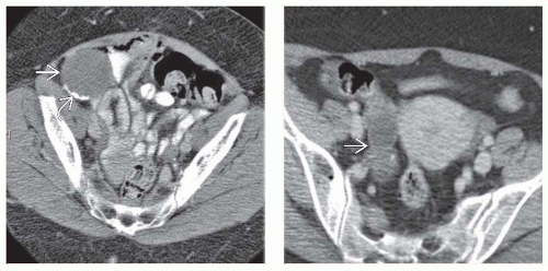 (Left) Axial CECT shows a cystic mass  in the right lower quadrant intimately associated with the cecum. There is some calcification in the right lower quadrant intimately associated with the cecum. There is some calcification  within the wall of the mass. (Right) Axial CECT shows a tubular, nonenhancing structure within the wall of the mass. (Right) Axial CECT shows a tubular, nonenhancing structure  arising from the tip of the cecum. There is no inflammatory infiltration or wall enhancement, as might be expected for acute appendicitis. arising from the tip of the cecum. There is no inflammatory infiltration or wall enhancement, as might be expected for acute appendicitis. |
Related posts:
Stay updated, free articles. Join our Telegram channel

Full access? Get Clinical Tree


