Chapter Outline
Bowel Wall: Pathologic Changes
Mesenteric and Omental Fat Disease
Multidetector computed tomography (MDCT) is currently the premier imaging technique for evaluating luminal, mural, and mesenteric abnormalities of the gastrointestinal tract. Although magnetic resonance (MR) enterography is rapidly gaining acceptance as the method of choice in evaluating patients with Crohn’s disease, MDCT examinations of the entire abdomen and pelvis can be acquired in seconds with near-isotropic voxels, allowing for high-quality and clinically useful volume imaging. The CT datasets can be viewed in any plane, and 3D techniques can be used to display large datasets effectively and graphically in a user-friendly format that is understandable to referring physicians.
Lumen Opacification
Proper distention and marking of the bowel lumen are vital in detecting mural thickening and excluding mural masses and mesenteric and omental disease. There are a number of methods available to accomplish this goal; the choice depends on the clinical setting.
In the emergent setting, in which bowel obstruction or intestinal ischemia is suspected, the intestinal secretions are usually sufficient to highlight the lumen, especially in a high-grade bowel obstruction. Orally administered contrast may be vomited and may remain in the stomach because of absent or diminished gastrointestinal motility. Finally, in cases of suspected ischemia, positive oral contrast media often hamper the creation of CT angiograms.
As part of a general survey examination of patients with no localizing signs or symptoms, a study performed with positive luminal contrast material is often obtained. As a caveat, the assessment of mural and mucosal enhancement of the gut may be compromised with the positive contrast material. Additionally, positive intraluminal contrast will interfere with CT angiography and some 3D techniques.
Air or carbon dioxide as an intraluminal contrast agent is used in the setting of CT gastroscopy and colonography.
Positive Contrast Agents
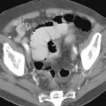
Positive contrast opacification (>75-100 HU) of the gut is accomplished by giving 1% to 2% barium suspensions or 2% to 3% solutions of iodinated water-soluble agents ( Fig. 5-1 ). The low percentage of barium requires commercial preparations made specifically for CT, in which additives are used to ensure that the barium remains in suspension.

In most patients, contrast material will reach the distal ileum within 45 minutes after initiation of drinking. Prolonged transit times are to be anticipated. Some common conditions altering the transit time include recent postoperative status, serum electrolyte disturbances, collagen vascular diseases (e.g., scleroderma), hypothyroidism, and intestinal obstruction. Conversely, patients who are hyperthyroid, have syndromes with associated increased intestinal motility (e.g., carcinoid, islet cell tumor), or have infections (e.g., cryptosporidiosis, giardiasis) will display a significantly accelerated intestinal transit time.
The choice between oral barium suspensions and water-soluble agents is dictated by the experience and preference of the radiologist. Water-soluble agents should be used exclusively for patients with abdominal trauma or a suspected perforated viscus, those who have a high likelihood of immediate surgery, and as an aid in percutaneous CT biopsy or other interventional procedures.
Neutral Contrast Agents
Neutral contrast agents (0-25 HU) have several advantages over positive contrast agents for evaluating mucosal, mural, and serosal disease ( Fig. 5-2 ). They allow excellent depiction of mural enhancement of the gut without the algorithm undershoot or overshoot that may accompany intraluminal high-attenuation, positive contrast, and low-attenuation gas. Neutral contrast agents also facilitate the performance of CT angiography and other three-dimensional techniques. Neutral contrast agents include water, milk, lactulose, 0.1% solution of barium (VoLumen; Bracco Diagnostic, Princeton, NJ), and water with mannitol or polyethylene glycol. Water can be administered as an effective neutral contrast agent for the upper gastrointestinal tract, especially the stomach and duodenum. However, it is less effective in distending the more distal bowel because it is normally absorbed before reaching the distal ileum.

These neutral agents are also helpful when performing CT angiography for staging and preoperative evaluation of hepatic, biliary, and pancreatic malignancies. Regardless of the agent, consistent opacification and/or distention of the jejunum remains a challenge. Unlike the standard small bowel series, periodic overhead films are not obtained before a CT. Thus it is impossible to determine the best time to scan in relation to proximal small bowel opacification and distention.
Gas Contrast
Gaseous distention of the stomach is important when evaluating mucosal and mural disease. It has been used with great success in CT gastrography for the diagnosis and staging of upper gastrointestinal malignancies.
For CT colonography, room air or carbon dioxide is insufflated per rectum ( Fig. 5-3 ). Adequate gaseous distention of the colon is very important for image interpretation because significant lesions may be obscured in a collapsed segment of colon. A complete discussion of this technique is presented in Chapter 53 .

Vascular Opacification
Opacification of the blood vessels is essential for complete evaluation of inflammatory, infectious, neoplastic, vascular, and traumatic diseases of the gastrointestinal tract. Obviously, this cannot be performed in all clinical settings (e.g., in cases of poor renal function or poor venous access). For general diagnostic cases, 100 to 150 mL (depending on concentration) of nonionic contrast is administered at a rate of 3 mL/s with a power injector. If CT angiography or other 3D techniques are to be performed, the rate is increased to 5 mL/s. Many sites use some form of bolus tracking as a method of timing the scan in relation to the level of arterial opacification.
One of the advantages of MDCT is that multiple datasets can be acquired with a single bolus of contrast. The following possible imaging times may be used when imaging the abdomen and pelvis—unenhanced, early arterial phase (20 seconds); late arterial-enteric phase (40 seconds); portal venous phase (70-90 seconds); equilibrium phase (210 seconds); and delayed phase (15-20 minutes).
For general survey abdominal imaging, obtaining scans during the portal venous phase is adequate. When assessing the viability of bowel, searching for a source of gastrointestinal hemorrhage, evaluating the cirrhotic liver, and searching for hypervascular metastases, it is useful to obtain noncontrast scans as well as scans during the later hepatic arterial and portal venous phases. When CT angiography is performed, scans should be obtained during the early arterial phase. The enteric phase corresponds to the late arterial phase and has been found useful for evaluating Crohn’s disease activity.
Normal Bowel Wall
Almost all significant pathology of the bowel wall results in mural thickening, which is often accompanied by changes in the attenuation of the bowel wall caused by edema, hemorrhage, tumor, fat, or gas. Two of the most common pitfalls in interpreting CT examinations of the gut are (1) confusing an insufficiently distended loop of bowel for pathologic thickening and (2) mistaking an inadequately opacified bowel loop for an abdominal mass. Techniques for achieving lumen distention are in the preceding section.
Esophagus
The esophagus has a length of 23 to 25 cm in the average adult. The wall of the distended esophagus is 3 mm in thickness ( Fig. 5-4 ). When the esophagus is not distended, the wall may approach 5 to 7 mm. It may be very difficult to differentiate diffuse and even focal esophageal disease if the lumen is not distended (a common occurrence). Furthermore, a hiatal hernia commonly has the appearance of focal wall thickening. The cervical esophagus lies posterior to the trachea in the midline. It may normally bulge into the posterior aspect of the trachea because of the limited space of the neck.

At the level of the thoracic inlet, the esophagus courses to the left of the midline and then lies adjacent to the left main stem bronchus and pericardium of the left atrium in the midthorax. More distally, the esophagus lies anterior to the descending aorta to the left of the midline as it enters the esophageal hiatus of the diaphragm. Normally, the thoracic esophagus should not indent the trachea, and there should be a triangle of fat between the aorta, spine, and esophagus distally. In patients with invasive esophageal carcinoma, the trachea is bowed along its posterior aspect by the esophageal tumor. More distally, invasive esophageal neoplasms obliterate the triangle of fat.
Contrast-enhanced examinations should show uniform mural enhancement of the esophagus, without mural stratification.
Stomach
The stomach is a functionally and anatomically dynamic organ, and its appearance depends on the degree of luminal distention and gastric location ( Fig. 5-5 ). For the well-distended, nondependent gastric fundus and body, a wall thickness of up to 5 mm is considered normal. The mural thickness of the antrum, however, is affected by anatomic and functional factors that make it normally thicker than other portions of the stomach. The gastric smooth muscle, particularly the circular layer, is thicker and denser in the antrum compared with more proximal portions of the stomach. Periodic concentric and eccentric antral contractions, as seen fluoroscopically, also contribute to the apparent mural thickening of the stomach.

When the normal stomach is distended with neutral contrast, enhancement of the mucosa may be seen, highlighted against the lower attenuation mucosa and muscularis propria. Up to 25% of patients show linear submucosal low attenuation or mural stratification in the antrum on contrast-enhanced MDCT examination. This may be partly the result of fat deposition in the submucosa.
Small Bowel
The normal small bowel is approximately 22 feet long and is suspended by a root that measures 6 to 9 inches and courses from the level of the ligament of Treitz caudally to the level of the ileocecal valve. As with conventional barium small bowel examinations, there are more valvulae conniventes in the jejunum than in the ileum. The normal small bowel wall measures between 1 and 2 mm when the lumen is well distended with a positive, neutral, or air contrast medium ( Fig. 5-6 ). When collapsed, the normal mural thickness of the small bowel measures between 2 and 3 mm.

The normal small bowel wall appears to have the greatest level of enhancement during the enteric phase (approximately 40 seconds, after initiation of the contrast injection). This investigation did not take into account the location of the small bowel when assessing bowel wall enhancement. Some investigators think that this is the ideal time to scan in patients with Crohn’s disease. Other investigators, using timed MR scanning after the injection of contrast, have shown that the maximal difference between normal and active inflammatory small bowel Crohn’s disease occurs much later, even several minutes after contrast injection. Furthermore, one investigation has shown that there is no significant difference in detecting the CT features of Crohn’s disease at 40 and 70 seconds postcontrast.
Regardless, the wall of collapsed segments of the small bowel has a greater attenuation than the wall of distended loops. Also, because the duodenum has more folds than the jejunum and the jejunum has more than the ileum, the duodenum enhances more than the jejunum and the jejunum enhances more than the ileum. Because collapsed small bowel loops have increased attenuation, similar to that of inflamed bowel loops, secondary findings of infectious or inflammatory small bowel disease (e.g., stratified enhancement pattern, engorged vasa recta, creeping fat of the mesentery, enlarged lymph nodes) should be considered (see Chapter 41 ).
Colon
The thickness of the colon wall as imaged on MDCT depends on the degree of distention. Fecal contents, fluid, colonic redundancy, and muscular hypertrophy (myochosis) make accurate determination of the true colonic wall thickness difficult. The normal colon wall (see Fig. 5-6 ) is normally less than 4 mm thick with proper distention. The normal wall is typically homogeneous in attenuation. With obesity becoming increasingly prevalent, submucosal fat is being identified in otherwise normal patients throughout the gut, but particularly in the colon.
Bowel Wall: Pathologic Changes
How to Examine the Bowel with MDCT
MDCT findings using thin section reconstructions (<1-2 mm), often overlapping, can be reconstructed in multiple planes. As in plain film radiography, orthogonal views are essential to assess the small bowel. We find that the axial and coronal planes are most helpful, with occasional sagittal reconstruction, especially if mesenteric artery occlusion is an important part of the investigation (i.e., ischemic bowel). Without orthogonal views, segments of the bowel may not be adequately assessed. Furthermore, in patients with Crohn’s disease, strictures and fistulas may not be identified. As an added benefit, surgeons and gastroenterologists are more familiar with viewing the small bowel in the coronal plane.
When viewing MDCT depictions of the bowel, especially the small bowel, we find that the routine scrolling on a workstation from superior to inferior and back, as well as anterior to posterior, allows the radiologist to follow bowel loops from the jejunum to ileum in a continuous fashion. Also, viewing the images in this volume fashion, changing windows and levels as needed, can detect mural abnormalities and distinguish the small bowel from mesenteric processes. Viewing the bowel with narrow windows, similar to liver windowing and leveling, can facilitate detection of hypervascular small bowel tumors, vascular abnormalities of the bowel wall (arteriovenous malformations and Dieulafoy lesions), and the mural hyperenhancement identified in active, inflammatory Crohn’s disease. Changing the window and level back to a soft tissue window after assessing the small bowel will facilitate identification of mesenteric abnormalities, which might have been missed using the narrow windowing and leveling. This dynamic volume interpretation is a demanding process but, once mastered on a modern interpreting workstation, can be performed rapidly and efficiently.
Evaluation of the Abnormal Gut
Mural thickening is the pathologic hallmark of gastrointestinal disease. When evaluating the abnormal gut, the following features should be carefully analyzed: mural attenuation and enhancement patterns ( Table 5-1 ); degree of mural thickening ( Table 5-2 ); symmetry of bowel wall thickening ( Table 5-3 ); and length of the diseased segment ( Table 5-4 ). Wittenberg and colleagues described a classification system for the abnormal bowel wall as depicted by MDCT ( Fig. 5-7 ). Regardless of enhancement patterns, symmetry of the bowel wall thickening, and length of the diseased segment, the thicker the bowel wall, the more likely that neoplastic disease is present. As a good rule of thumb, confirmed in the literature as well as anecdotally, wall thickening of 1.5 cm or less is infectious and/or inflammatory and wall thickening more than 1.5 cm is neoplastic. Between 1 and 2 cm, there is overlap between the categories. Additionally, neoplastic disease tends to cause asymmetric wall thickening, but this general rule holds true for over 90% of cases.
|
Stay updated, free articles. Join our Telegram channel

Full access? Get Clinical Tree


