Multiple masses. Sigh. Do you hate these? Take a deep breath and lean into it. These masses are really not that hard to assess. They are almost always benign, BI-RADS 2, representing multiple cysts or fibroadenomas. This is like a “Get Out of Jail Free” card in Monopoly. Before we use the card though, we need to carefully look at each one to ensure that a cancer is not masquerading as a benign mass. We’ll give you our best tips for figuring out which patients actually need recall and which can be seen next year. Soon you will be thinking, “Bring it on!” instead of putting these cases on your partner’s desk.
The rule of multiplicity in mammography is that multiple and bilateral similar-appearing findings with low-suspicion features have a very high probability of being benign. The basis for this rule is that the genetic, hormonal, and other influences that produce benign findings in one breast tend to produce similar findings in the other, and it is very uncommon for malignant lesions to present simultaneously as similar-appearing findings in multiple regions of both breasts.
When multiple bilateral masses with circumscribed margins are detected by mammography, the rule of multiplicity can be applied and the findings are considered benign. These women can be spared the discomfort, inconvenience, anxiety, and cost of diagnostic mammography, ultrasonography (US), and in some cases, aspiration or biopsy.
However, with multiple masses, it is important to avoid the pitfall of using the rule of multiplicity too soon. Don’t play your “Get Out of Jail Free” card too early. These cases are complex and can be difficult to interpret. Benign masses may obscure other lesions or distract the radiologist from findings that need further evaluation.
Etiology of Multiple Bilateral Masses
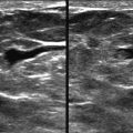
Multiple bilateral masses may represent one type of lesion or more than one type. Cysts are the most common lesions contributing to multiple bilateral masses ( Figs. 8-1 and 8-2 ). Cysts can develop at any age, but are most common in premenopausal women. Milk of calcium may be seen mammographically, providing clues to the cystic nature of one or more of the masses.


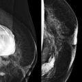
Fibroadenomas are the second most common lesion causing multiple bilateral masses. When coarse calcifications are seen within one or more of the masses, it suggests that other, noncalcified masses may represent fibroadenomas as well ( Fig. 8-3 ).

Peripheral papillomas uncommonly present as multiple bilateral masses. Multiple papillomas are usually round or oval masses with circumscribed margins and are associated with an increased risk for breast cancer. Papillary carcinoma commonly presents as multiple circumscribed masses with segmental distributions on mammography.
Malignant multiple bilateral masses are very uncommon. Nevertheless, metastatic disease should still cross your mind when evaluating multiple bilateral breast masses ( Fig. 8-4 ). Most women with breast metastases have a known history of malignancy. Unfortunately, breast involvement may be the first sign of recurrent disease in a woman thought cured of a malignant disease. Melanoma is the most common nonbreast primary neoplasm to metastasize to the breast. The appearance of the mammogram is usually different from that seen in the benign causes of multiple masses. The masses are typically denser than cysts or fibroadenomas, and the margins are often ill defined. Metastatic lesions of the breast are typically located throughout the organ, including the fatty tissue in the inframammary fold and axilla, whereas cysts and fibroadenomas are located within the breast tissue. Metastatic disease to the breast is often associated with axillary adenopathy, and some of the breast masses may represent metastatic intramammary lymph nodes. Lymphoma, leukemia, and rhabdomyosarcoma are rare in the breast and will be covered in more detail in Chapter 11, Expanding the Differential Diagnosis .

Intramammary lymph nodes typically demonstrate a lucent notch representing the fatty hilum. However, if the lucent notch is not visible, lymph nodes may contribute to the pattern of multiple bilateral masses. When lymph nodes have a characteristically benign appearance, we do not consider them one of the multiple bilateral masses. For example, if there are two indeterminate circumscribed masses in one breast and a typical intramammary lymph node in the other, the findings are not considered as multiple bilateral masses.
Skin lesions, such as nevi or neurofibromas, can mimic multiple bilateral breast masses. Ideally, your technologists will help you by noting them on the clinical history sheet ( Box 8-1 ).
- •
Cysts
- •
Fibroadenomas
- •
Papillomas
- •
Metastatic disease or lymphoma (really uncommon!)
- •
Skin lesions (mimic)
Management of Multiple Bilateral Masses
“Multiple bilateral masses” refers to asymptomatic women whose mammograms show a minimum of three masses, with at least one mass in each breast. These findings occur in 0.5% to 1.7% of patients in a screening population.
Management recommendations for multiple bilateral masses with benign features differ among radiologists. Many assign BI-RADS 2 and recommend annual screening mammography. Some radiologists recommend bilateral US after the initial mammogram, to serve as a baseline for future studies, while others assign BI-RADS 3 on the initial mammogram and recommend a 6-month bilateral follow-up study to evaluate stability. Our preference is to assign BI-RADS 2 and recommend annual mammographic screening. However, if any of the masses has suspicious features, we have a low threshold for recalling the patient for targeted diagnostic evaluation, as we demonstrate in some of the cases in this chapter.
Why not bring them all in for US? The use of US to evaluate multiple bilateral masses with benign mammographic features yields frequent false-positive findings. As a group, these women do not appear to be at increased risk for developing breast cancer. In women with fibrocystic changes, complicated cysts or complex masses are frequently detected. Benign solid masses, which also frequently occur in these patients, often have indeterminate sonographic features. A degenerating fibroadenoma may have shadowing on US. When masses other than simple cysts are identified by US, they often prompt a recommendation for aspiration, biopsy, or short-term follow-up studies. In addition, when a mass is seen by US, it may be difficult or impossible to identify a corresponding mammographic finding with certainty, unless a localizing intervention is performed.
What Is the Evidence?
There is strong support in the literature, when certain criteria are met, to consider multiple bilateral masses benign, even when there are no previous mammograms for comparison. studied 1440 women with multiple bilateral masses, with circumscribed margins, whose mammograms were prospectively interpreted as benign, irrespective of the availability of prior mammograms. In fact, no comparison studies were available in almost half of the cases. The interval cancer rate for women with multiple bilateral masses was 0.14%, which is similar to previously reported interval cancer rates for the general population and lower than the incident cancer rate in the United States. In an earlier study ( ), 1 cancer was found at follow-up of 253 women (0.4%) with multiple bilateral well-defined solid masses. In the ACRIN 6666 trial, none of 39 women with multiple bilateral circumscribed masses on mammography had cancer. Likewise, none of 48 women with multiple solid masses with benign features on whole breast US had cancer.
Criteria for Assigning BI-RADS 2 Category to Multiple Masses
The Masses Must Be Bilateral
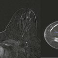
Multiple circumscribed masses in one breast do not fulfill the multiplicity rule and cannot be considered benign at screening. Unilateral masses with benign features are still most likely to represent cysts or fibroadenomas, and sometimes papillomas. However, the risk of carcinoma is higher than when the masses are bilateral ( Fig. 8-5 ). Suspicion for malignancy is increased if the masses are in a segmental distribution.

The Masses Must Be Multiple
Because the masses must be bilateral, the minimum number of masses present is three; two in one breast and one in the other breast.
The Margins Must Be Circumscribed
A circumscribed margin is a benign feature. When multiple masses are present, some of the mass margins may be obscured. The BI-RADS lexicon defines “circumscribed” as having at least 75% of the margin visible as circumscribed. If a mass is not obscured and most of the mass is circumscribed but the remainder of the margin is ill defined, irregular, or spiculated, then diagnostic evaluation is needed.
Evaluating Multiple Bilateral Masses
The presence of multiple bilateral masses can often be appreciated on an overview of the case images. Are you finished? Sorry, but no. You can’t use the “Get Out of Jail Free” card until you really look! There is no evidence that the presence of multiple bilateral circumscribed masses reduces the chance that a malignant lesion will also develop. To find her cancer, look for mass features that distinguish one from the others. Next, ignore all those masses and look at everything else. Last, be aware of new or changing clinical findings described by the patient or her health care provider.
Which One of These Is Not Like the Others?
Do you remember playing that game when you were a kid? You had to figure out which of several very similar objects was different. We’ll bet that you were really good at that game because you are a radiologist now!
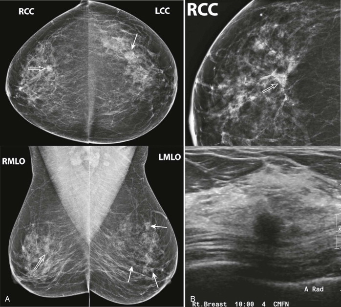
When multiple bilateral masses are present, their size, shape, density, and margins should be individually examined. Is one mass substantially larger than the others (dominant)? Search for masses that stand out from others, such as those that are very dense, irregular in shape, or have indistinct, microlobulated, or spiculated margins ( Figs. 8-6 to 8-8 , and Box 8-2 ). Search for associated suspicious findings such as calcifications, architectural distortion, adenopathy, skin thickening, or nipple retraction.