Muscle and Nerve on FDG PET/CT


Abstract


Muscles and nerves may demonstrate benign physiologic FDG avidity and must be distinguished from malignancy. This chapter describes common appearances of benign FDG avidity in muscles and nerves and compares them to the appearance of malignancy.


Keywords

FDG, PET/CT, muscle, nerve


Muscles are a less common site of malignancy, yet are very important to the proper interpretation of 18F-flourodeoxyglucose (FDG) positron emission tomography/computed tomography (PET/CT) in their own right. Benign skeletal muscle may demonstrate substantial FDG uptake, and this should not be confused with malignancy. Physiologic FDG avidity in skeletal muscle is often related with muscular exertion, which is why at most FDG PET facilities patients are asked to refrain from exercise the day before and the day of a PET/CT scan. Linear FDG avidity corresponding to normal appearing muscles on CT is usually physiologic ( Fig. 4.1 ).


FIG. 4.1


Physiologic Muscular FDG Avidity.

(A) Axial fused PET/CT and CT images, as well as (B) sagittal CT and fused PET/CT images, demonstrate physiologic FDG avidity within the bilateral rectus abdominus muscles (arrows) , in a patient who performed sit-ups the morning of FDG PET/CT scan, despite instructions not to exercise before the examination.


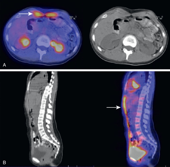
Extensive physiologic FDG avidity could initially be alarming on PET/CT examinations. For example, large segments of FDG-avid back musculature are uncommon and could mimic an aggressive process. However, correlation with the corresponding CT images usually allows for the proper identification of physiologic muscular FDG avidity. Note the patient in Fig. 4.2 . This patient has extensive FDG avidity in the region of back musculature. The corresponding CT images demonstrate normal fat planes within the FDG-avid muscle, consistent with benign muscle, despite the extensive FDG avidity. FDG-avid masses would be expected to displace or obliterate the normal fat planes within musculature ( Fig. 4.3 ). While areas of FDG-avid malignancy underlying the physiologic muscular FDG avidity cannot be entirely excluded, the presence of normal fat planes within musculature is a very reassuring sign that FDG avidity is probably physiologic.


FIG. 4.2


Extensive Physiologic Muscular FDG Avidity.

(A) Maximum intensity projection (MIP) PET demonstrates extensive FDG-avid musculature, including bilateral FDG avid paraspinal musculature (arrows) . (B) Sagittal CT and fused PET/CT images demonstrate the FDG avidity in paraspinal musculature (arrow) . The CT image demonstrates normal fat planes (arrowheads) traversing the FDG-avid muscles, a reassuring sign that the FDG avidity is probably physiologic.


FIG. 4.3


FDG-avid Muscular Lymphoma.

(A) MIP PET demonstrates midline FDG avidity overlying the chest and upper abdomen (arrow) , as well as additional lesions osseous foci (arrowhead) . (B) Axial CT and fused PET/CT images demonstrate the midline FDG avidity localizes to paraspinal musculature (arrow) . The CT image demonstrates obliteration of normal fat planes in the region of the FDG avidity (arrow) , as compared with the preservation of fat planes in the contralateral muscle (arrowhead) . The obliteration of fat planes is worrisome for a pathologic process, and biopsy demonstrated lymphomatosis infiltration of the muscle.


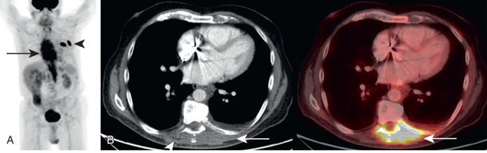
As with the skeleton, FDG PET helps visualize metastases that may be occult or overlooked on anatomic imaging. Muscle is an uncommon site for metastasis; therefore it is an organ that may not be scrutinized by the radiologist on every scan. FDG avidity highlights potentially important findings for additional scrutiny ( Fig. 4.4 ).


FIG. 4.4


FDG-avid Muscular Metastasis.

Axial CT and fused PET/CT images demonstrate an FDG-avid focus in the left rectus muscle (arrow) . Corresponding CT image demonstrates a subtle enhancing lesion at this location (arrowhead) , suspicious for metastasis, which could easily have been overlooked without FDG guidance. Biopsy confirmed a muscular metastasis.


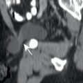
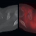
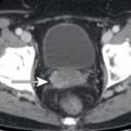
Unfortunately, as with other organ systems, FDG avidity could lead to the misinterpretation of benign findings. Focal areas of physiologic muscular FDG avidity could be mistaken for FDG-avid lymph nodes or other suspicious FDG-avid lesions. One area of muscle for which mistakes may occur is the crura of the diaphragm. The diaphragmatic crura may appear to have focal FDG avidity on individual axial slices, and this may be confused with retrocrural or upper abdominal lymph nodes ( Fig. 4.5 ). Similarly, not all histologies of malignancy will be readily apparent on FDG PET, and the corresponding CT images may demonstrate an abnormality within the muscle better than the FDG PET images. For example, myxoid tumors will demonstrate low attenuation compared to skeletal muscle, and muscular metastases from myxoid tumors may be more readily apparent on the CT component of the PET/CT ( Fig. 4.6 ).


FIG. 4.5


Benign FDG Avidity in the Diaphragm, Which Could be Mistaken for Malignancy.

Axial PET, CT, and fused PET/CT images demonstrate FDG-avid foci in the midline upper abdomen (arrows) as well as in the region of the left costodiaphragmatic recess (arrowhead) . The midline foci could be confused with retrocrural or upper abdominal lymph nodes, while the left costodiaphragmatic foci could be confused with pleural metastases. The CT and fused images demonstrate that the midline foci localize to normal-appearing diaphragmatic crura, while the left costodiaphragmatic foci localize to the lateral pleura, without any nodal or pleural masses.


FIG. 4.6


Muscular Metastasis from Myxoid Liposarcoma More Readily Apparent on CT Than FDG PET Images.

Axial PET, CT, and fused PET/CT images demonstrate minimal FDG avidity in right thigh musculature. The CT image demonstrates a low-attenuation mass in the musculature with faint areas of enhancement (arrows) . The mild FDG avidity was not mentioned in the FDG PET/CT report, probably overlooked as physiologic muscular FDG avidity. The abnormality was mentioned in a separate contrast-enhanced CT report. Biopsy confirmed muscular metastasis. This case highlights the importance of evaluating CT images to detect non- or mildly FDG-avid malignancy. This case also highlights the importance of knowing the tumor histology. Myxoid tumors often produce low-attenuation masses, leading the radiologist to carefully evaluate for low-attenuation lesions.

Only gold members can continue reading. Log In or Register to continue

Stay updated, free articles. Join our Telegram channel

Jun 18, 2019 | Posted by in GENERAL RADIOLOGY | Comments Off on Muscle and Nerve on FDG PET/CT

Full access? Get Clinical Tree

Get Clinical Tree app for offline access