, Stefano Fanti1 and Lucia Zanoni1
(1)
Department of Nuclear Medicine, Universitary Hospital Sant’Orsola-Malpighi, Bologna, Italy
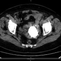
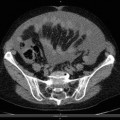
Osteoporotic Fractures of the Pelvis
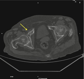
Fig. 6.1
Fracture of the right iliac-pubic ramus
Pelvic fractures encompass a broad spectrum of injuries, from low-energy osteoporotic fractures to high-energy disruptions of the pelvic ring. Following low-energy trauma fractures are frequently classified as pelvic girdle fractures. CT is both sensitive and specific, and it is a valid alternative to MRI in localizing the fracture line. The CT scan aspect varies depending on the degree of fracture healing; indeed it can highlight sclerotic healing or fresh fracture lines. Important issue is to rule out malignancy and osteomyelitis.
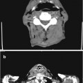
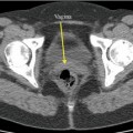
Osteoid Osteoma
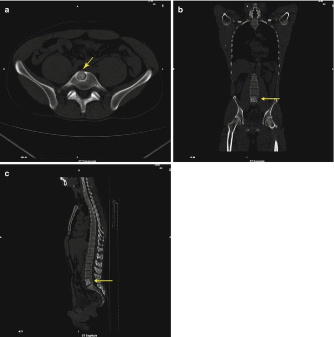
Fig. 6.2
Non-avid round lesion in L5 vertebral body: (a) axial, (b) coronal, and (c) sagittal views (arrows)
Osteoid osteoma is a small benign tumor of bone that is usually present at a young age and causes moderate-to-severe bouts of intermittent pain, often worse at night but partially relieved by non-steroidal anti-inflammatory drugs. It appears as a circumscribed subcortical annular lesion with a double-attenuating sign with thin curvilinear or serpiginous low-density grooves related with enlarged feeding arterioles (“the vascular groove sign”). It is easy to visualize a central nidus among the surrounding dense bone sclerosis (periosteal reaction). CT scan is particularly helpful to define the precise localization of the nidus in the hip or spine.
Lipoma
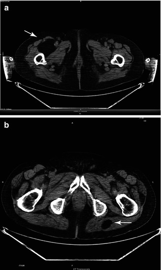
Fig. 6.3
Low-density mass in the right thigh (a) or in the left gluteus (b)
Lipomas arising from fat in the intramuscular septa cause a diffuse, palpable swelling, which is more prominent when the related muscle is contracted. Lipoma is a slow-growing, benign fatty tumor form characterized by soft, lobulated mass enclosed by a thin, fibrous capsule. There is low-density tissue (typically approximately −65 to −120 HU), identical to subcutaneous adipose tissue. Thin septa can be found.
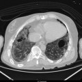
Extramedullary Hematopoiesis (EMH)
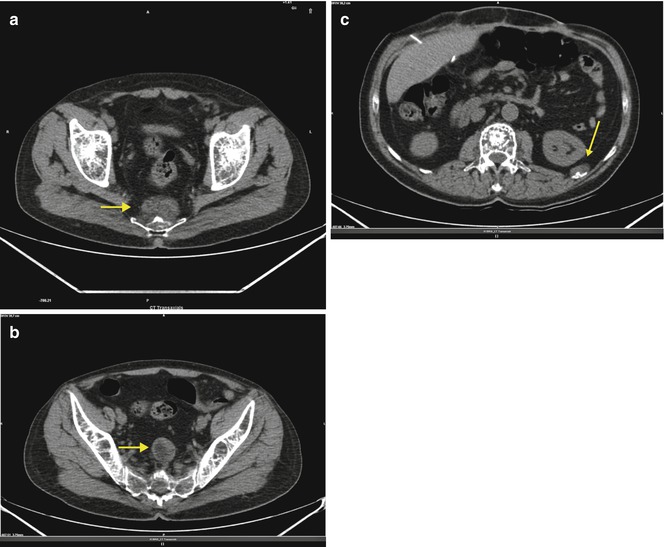
Fig. 6.4
(a) Pre-coccyx (arrow), (b) presacral (arrow), and (c) thoracic tumorlike mass adjacent to a left rib (arrow)
EMH is a response to erythropoiesis failure in bone marrow. It is one of the common features of chronic myeloproliferative disorders. The neoplastic stem cells have the capacity to circulate and migrate to secondary hematopoietic organs (mainly spleen and liver and occasionally lymph nodes or other organs) causing macroscopically visible tumorlike masses. The lesions are usually round masses with lobulated margins, they don’t calcify and do not usually cause bone erosion, and they appear of low attenuation on non-contrast CT. In the thorax they can be visible with widening of the ribs or around the expanded posterior or anterior part of the ribs: the marrow overflows from the bone, destroys the cortex (however, the cortex can be found intact), and spreads around it. A similar process may start form a vertebral body.
Vertebral Collapse
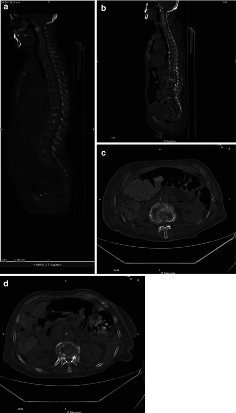
Fig. 6.5
(a–d) Initial non-traumatic vertebral collapse sagittal (a, b) and axial (c, d) views
The following CT findings are significantly more frequent in benign (osteoporotic) vertebral collapse: cortical fractures of the vertebral body without cortical bone destruction, retropulsion of a bone fragment of the posterior cortex of the vertebral body into the spinal canal, fracture lines within the cancellous bone of the vertebral body, an intravertebral vacuum phenomenon, and a thin diffuse paraspinal soft-tissue mass.
On the contrary the following CT findings are significantly more frequent in malignant collapse (metastatic or myelomatous): destruction of the anterolateral or posterior cortical bone of the vertebral body, destruction of the cancellous bone of the vertebral body, destruction of a vertebral pedicle, a focal paraspinal soft-tissue mass, and an epidural mass.
Spina Bifida
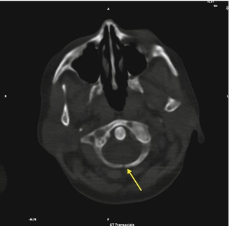
Fig. 6.6
Non-fused posterior arch of C1 (arrow)
Related posts:

Stay updated, free articles. Join our Telegram channel

Full access? Get Clinical Tree


