My Aunt Minnie
Amorphous calcifications
Artifacts
Calcified parasites
Cysts
Dystrophic calcifications
Extracapsular implant rupture
Fibroadenolipomas
Gel bleed
Hair
Hickman catheter
Hyalinizing fibroadenomas
Implants
Intracapsular implant rupture
Keloids
Lipomas
Lucent centered calcifications
Lymph nodes
Milk of calcium
Negative density artifacts
Nipple rings
Oil cysts
Plus density artifacts
Radiolucent mass
Rod-like calcifications
Seborrheic keratoses
Skin folds
Sternalis muscle
Vascular calcifications
Wire fragments
Wire localization
The term “Aunt Minnie” is used in radiology to characterize lesions that have a distinctive, unique appearance. Most radiology residents learn about Aunt Minnie early in their careers. While planning this book, I thought a chapter on Aunt Minnie would be easy to put together. I have discovered that this is not so! At least in mammography, the concept of Aunt Minnie is difficult to apply and I have struggled in selecting what should be included in this chapter. Does Aunt Minnie really always look the same? If for the same entity there is some variation in appearance, can it still be Aunt Minnie? Is your Aunt Minnie the same as my Aunt Minnie?
By this point you are probably wondering why I am dwelling on this. Probably this is by way of a disclaimer! I have elected to illustrate entities I define as Aunt Minnie. Some of you may not recognize my Aunt Minnie; however, the entities presented are distinctive and should be recognized as benign or iatrogenic. Rarely do these require additional evaluation, short-interval follow-up, or intervention.
In approaching “patient” (as opposed to “case”) discussions, consider the 4 D’s. The first is detection. Is there a potential abnormality? The second is a description of a confirmed finding based on complete information (e.g., additional clinical and imaging evaluations). Your description should lead you and the listener to a differential and the likely diagnosis you will propose. The third D is your differential. When considering the possibilities, remember that all findings have benign and malignant considerations and you should move through the list in a logical manner. Try not to jump back and forth from benign to malignant. Tell the listener what you think the lesion could be, not what it is not. Also, try to be specific; saying that “this is likely a malignancy or cancer” is not very insightful. The last D is what you think the diagnosis is most likely to be.
Patient 1
What is your diagnosis?
If you are not sure, what can you do to be 100% sure?
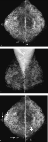
Multiple masses are projecting on the breast parenchyma bilaterally. On close inspection, air is seen as a thin radiolucency, partially or completely outlining the margins of several of the masses (Fig. 1.1C, thicker arrows). Those in tangent to the x-ray beam are seen extending beyond the breast (Fig. 1.1C, thin arrows). Bilateral skin lesions may be seen in otherwise healthy women or, when this numerous, in women with neurofibromatosis. These patients can be challenging. We are often mesmerized by benign findings and neg lect more subtle findings that potentially reflect breast cancer. So, do not let benign, obviously malignant, or clinical findings distract you from reviewing the mammogram completely. You need to be even more focused in looking for potential signs of early breast cancer around and in between obvious findings. If there is any question about a mass being on the skin you can examine the patient and, if still not sure, place a metallic BB on the identified skin lesion and obtain follow-up images with the BB (and skin lesion) in tangent to the x-ray beam.
BI-RADS® category 1: negative. BI-RADS® category 2: benign finding is used if the skin lesions are described in the body of the report. Next screening mammography is recommended in 1 year.
Patient 2
What is the diagnosis in these three patients, and how can you be certain?
What else could you do?
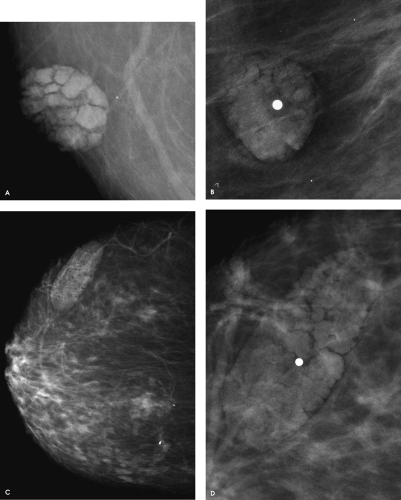
Seborrheic keratoses: Characteristic mammographic appearance is demonstrated with these three patients. When superimposed on the breast parenchyma, skin lesions are often partially, or completely, outlined by a thin radiolucency (air), as are the interstices of the verrucous lesions. When they are in tangent to the x-ray beam, their extension beyond the skin is outlined by air (radiolucent). Although metallic BBs are often used to mark these skin lesions, the mammographic appearance of the verrucous lesions is distinctive. Metallic BBs are more helpful on smooth skin lesions, because these are more likely to simulate a breast lesion when superimposed on the breast parenchyma on the two standard views of the breast (e.g., craniocaudal and mediolateral oblique views). When a technologist uses a BB, she indicates the reason on the woman’s history form and affixes a sticker on the films (e.g., “BB on mole” or “BB on lump”) indicating the reason for the use of the BB. Unless the skin lesions are too numerous to mark, the routine use of BBs to mark skin lesions can avert some callbacks.
Seborrheic keratoses are common benign epidermal tumors distributed on skin that bears hair. They do not develop on mucosal surfaces and are infrequent under the age of 30 years. They are common, typically multiple, and continue to develop as patients age. A familial predilection with a possible autosomal dominant form of inheritance has been described. Seborrheic keratoses are typically flat when they first develop and over time can become more verrucous, polypoid, and pedunculated in appearance. Rarely, rapid proliferation or increases in the size of pre-existing lesions may be an indication of an internal malignancy (Leser-Trélat sign).
Patient 3
What are imaging findings of intramammary lymph nodes?
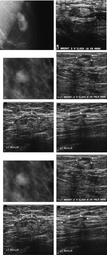
The mammographic appearance of lymph nodes is variable (Fig. 1.3A, C). Most commonly, intramammary lymph nodes are well circumscribed, oval (reniform), mixed-density (fat containing) masses localized to the upper outer quadrants. However, they can be found anywhere, including the inner quadrants, and may fluctuate in size and density. They may have a prominent fatty component relative to the water-density portion or vice versa. Rarely, lymph nodes disappear, only to reappear on subsequent mammograms. The mammographic appearance of “normal” lymph nodes is usually distinctive enough that no additional imaging is indicated.
Ultrasound is used adjunctively in patients in whom a lymph node is suspected but a fatty hilum is not definitely seen mammographically. Ultrasound is also the primary imaging modality used in patients who are under the age of 30 years, pregnant, or lactating, who present with a palpable finding. On ultrasound, lymph nodes are typically well-circumscribed masses with a hypoechoic cortical area and a hyperechoic central, or eccentric focus, corresponding to the fatty hilar region seen mammographically (Fig. 1.3B, D, E, F, G, H). If power Doppler is used, blood flow is seen associated with the hyperechoic region.
On T2-weighted magnetic resonance images, lymph nodes demonstrate high signal intensity. Following contrast administration, lymph nodes are characterized by rapid contrast uptake on
T1-weighted images. This uptake is followed by either a plateau or rapid washout. Contrast enhancement may appear rimlike when the hilar region is centrally located (Fig. 1.3I, J). Vessels can sometimes be seen in the hilar region.
Mammographically, if a lymph node increases in size and density and there is associated loss of the fatty hilum with indistinct or spiculated margins, biopsy may be indicated; in many women, however, these changes are reactive and do not reflect a malignant process. Similarly, on ultrasound, biopsy is considered if there is thickening and bulging of the cortical area, often asymmetric and microlobulated, with concomitant thinning and apparent mass effect, or complete loss, of the hyperechoic hilar region. Increased blood flow can be seen in some of these lymph nodes.
Patient 4
How would you describe the findings?
What is your diagnosis?
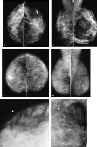
Mixed-density (fat containing) masses are imaged mammographically in these two patients consistent with fibroadenolipomas (also called hamartoma, breast-within-a-breast). Fatty, glandular and fibrous tissues are surrounded, and separated, from the remainder of the breast tissue by a fibrous pseudocapsule. As illustrated by these two patients, the proportions of each tissue type vary from patient to patient. In some women the lesions are mostly fatty, in others glandular tissue predominates. They can be detected on screening studies (Fig. 1.4A, B, left breast) or, in some patients can present as a palpable finding (Fig. 1.4C, D, right breast). Rarely, they occur in accessory axillary glandular tissue and may enlarge rapidly.
On ultrasound, fibroadenolipomas are usually separable from surrounding normal tissue and characterized by an admixture of hypo- and hyperechoic areas with disruption of normal tissue architecture. The posterior acoustic features of these lesions are variable and include no posterior acoustic features, enhancement, shadowing, or a combined pattern (areas of enhancement and areas of shadowing).
Variable combinations of adipose tissue, fibrous stroma, and lobular structures are seen histologically and, although separable from the adjacent breast tissue, hamartomas lack a true capsule. Rarely, myxoid and chondroid hamartomas are reported histologically when the lesions contain muscle and cartilage, respectively. A myxoid hamartoma has been reported in a 36-year-old male presenting with a slowly growing breast mass. Breast cancer can arise in fibroadenolipomas, so the tissue in these lesions should be evaluated for the development of any mass, distortion, or calcifications as thoroughly as breast tissue anywhere else.
BI-RADS® category 2: benign finding. Next screening mammogram is recommended in 1 year.
Patient 5
How would you describe the findings?
What is your diagnosis?
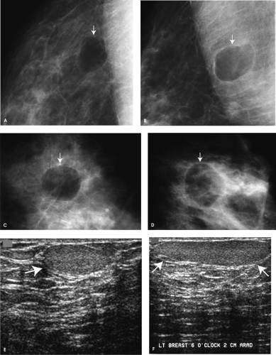
Radiolucent, well-circumscribed masses (small arrows) with a thin fibrous capsule consistent with lipomas; these rarely calcify. Unlike oil cysts, which have a variable sonographic appearance but often simulate cysts, lipomas are well circumscribed, solid, slightly hypo- or isoechoic masses on ultrasound (large arrows). In some women, lipomas can be slightly hyperechoic and a small amount of posterior acoustic enhancement may be noted (Fig. 1.5E, F). A gentle mass effect on surrounding tissue and Cooper ligaments can also be seen with some lipomas. Rarely, lipomas can be seen within the pectoral muscle.
BI-RADS® category 2: benign finding. Next screening mammography is recommended in 1 year.
Patient 6
How would you describe the findings?
What is your diagnosis?
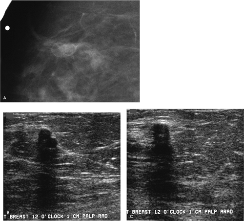
Two adjacent 5-mm round, well-circumscribed radiolucent masses are seen mammographically, corresponding to the “lump” described by the patient. Given the history of a reduction mammoplasty, these are consistent with oil cysts. The diagnosis is established mammographically. Radiolucent masses in the breast are benign. The benign nature of the palpable finding is discussed with the patient, and she is reassured that this is not breast cancer and that it will not become cancerous. A definitive report is issued to avoid unnecessary surgery.
BI-RADS® category 2: benign finding. Next screening mammography is recommended in 1 year.
On physical examination, there is a discrete, hard, lobulated mass palpated at the 12 o’clock position, 1 cm from the right nipple. A macrolobulated, nearly anechoic mass with significant shadowing is imaged, corresponding to the palpable finding. Although the ultrasound is included for illustrative purposes, the diagnosis is made on the mammographic findings (i.e., ultrasound is not indicated when a radiolucent mass is seen mammographically). Oil cysts can have a variable appearance on ultrasound, ranging from simulating a simple cyst to a complex cystic mass or a mass with significant shadowing as seen here.
Patient 7
How would you describe the findings?
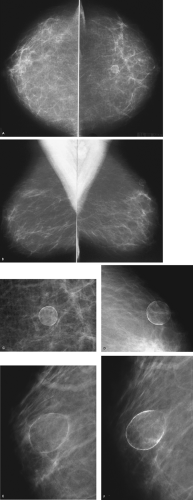
These are oil cysts with eggshell or rim calcification. Oil cysts commonly develop following trauma or surgery; however, most patients do not recall the trauma (and may not recall surgery). These are round or oval, well-circumscribed, radiolucent masses. As illustrated here, thin calcifications can develop in the wall of the cyst. With time these may stabilize or decrease progressively in size (Fig. 1.7E, F) and, in some patients, eventually resolve completely.
Steatocystoma multiplex is a rare, autosomal dominant condition characterized by the presence of multiple cutaneous cysts appearing during adolescence and increasing progressively with age involving the anterior trunk, back, proximal extremities, and external genitalia. Multiple oil cysts are seen mammographically.
BI-RADS® category 1: negative. BI-RADS® category 2: benign finding is used if the findings are described in the body of the report. Next screening mammogram is recommended in 1 year.
Patient 8
How would you describe the findings?
What procedure have both of these patients undergone?
A radiolucent mass with mural and internal curvilinear calcifications is seen in the left breast in Fig. 1.8A–C. The appearance of some of the internal calcifications suggests the presence of smaller lucent masses within the dominant mass. Also noted is asymmetric tissue inferiorly on the right mediolateral oblique view demonstrating a swirling pattern (Fig. 1.8B). The left breast is smaller than the right. The findings suggest a history of reduction mammoplasty, which is confirmed on the patient’s history sheet.
In the second patient, multiple mixed-density masses of varying sizes are present bilaterally (Fig. 1.8D). A few dense calcifications are associated with some of the masses (Fig. 1.8D, F). A year later, many more coarse calcifications are noted associated with the mixed-density (fat containing) masses (Fig. 1.8E). The calcifications are now more curvilinear in appearance, seemingly outlining a cluster of lucent masses (Fig. 1.8G). As the calcifications have increased, the overall size and associated soft tissue component of some of the masses has decreased. The presence of multiple, bilateral oil cysts may reflect a history of trauma or, as in this patient, reduction mammoplasty. Comparison films, if available, will be helpful in establishing a change in overall breast size following the surgery, as well as the development of the oil cysts.
What are the imaging findings associated with fat necrosis?
In the acute setting, fat necrosis following reduction mammoplasty may present with one or multiple, mixed-density masses; some may be spiculated. As the inflammatory process associated with fat necrosis resolves, single or multiple, uni- or bilateral oil cysts of varying sizes may remain. Although some may develop rim calcifications, most develop coarser, curvilinear calcifications, as demonstrated in these two patients. With time, these may stabilize, continue to calcify, or some eventually resolve completely. In some patients, these become palpable as they calcify 1 or 2 years following the surgery. When they are palpable, it is important to reassure the patient that the palpable finding is benign and requires no further intervention. It is also important that a definitive report be issued.
BI-RADS® category 1: negative. BI-RADS® category 2: benign finding is used if the findings are described in the body of the report. Next screening mammogram is recommended in 1 year.
Patient 9
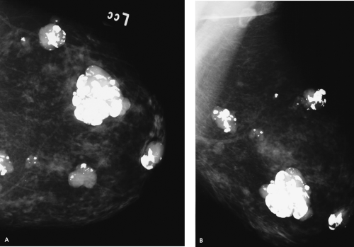 Figure 1.9. Screening study, 70-year-old woman. Craniocaudal (A) and mediolateral oblique (B) views, left breast. |
What is the diagnosis and what BI-RADS® category would you assign?
Multiple, well-circumscribed, dense masses, some with macrolobulations and varying amounts of associated dense, coarse calcifications, are present in the left breast. These are hyalinizing fibroadenomas with associated dystrophic calcifications (i.e., popcorn-type calcifications). As estrogen levels decrease, the epithelial component in fibroadenomas atrophies and is replaced by dense, hyalinized fibrous tissue. These changes may be characterized mammographically by a decrease in size, an increase in density, and the development of dense, dystrophic calcifications in a pre-existing well-circumscribed mass. Because these calcifications develop in hyalinized fibrous tissue, they are variable in size, shape, and density (i.e., there is no preformed space molding the developing calcifications). Also noted is a well-circumscribed, mixed-density oval mass superimposed on the pectoral muscle on the mediolateral oblique view, consistent with a lymph node.
BI-RADS® category 2: benign finding. Next screening mammogram is recommended in 1 year.
Patient 10
What observations can you make, and do you think this woman needs to be called back for additional evaluation?
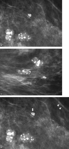
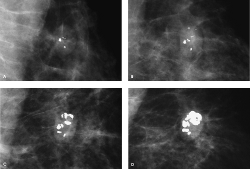
Two macrolobulated, dense masses with partially well circumscribed margins and coarse, dense calcifications are imaged in the left breast. A cluster of dense, tightly packed calcifications is also present (Fig. 1.10C, arrow), with no associated soft tissue component. Scattered benign calcifications and arterial calcification Fig. 1.10C arrowheads) are incidentally noted. These are all hyalinizing fibroadenomas. It is important to recognize that a mass is not always seen in association with the dystrophic calcifications of a fibroadenoma (i.e., a cluster of calcifications alone can represent a hyalinizing fibroadenoma). These findings require no additional evaluation or short-interval follow-up. However, don’t be lulled by obviously benign findings; make sure to focus your attention on the remainder of the mammogram.
Patient 11
What is your diagnosis?
An oval mass, with partially obscured and well-circumscribed margins and associated dense coarse calcifications, is imaged anterior to the pectoral muscle. On subsequent screening studies, the mass decreases slightly in size, becomes denser, and the calcifications increase in number, size, and density. These findings are diagnostic of a hyalinizing fibroadenoma with dystrophic calcifications (i.e., developing popcorn calcification). This finding is benign and requires no additional evaluation or short-interval follow-up.
Patient 12
How would you describe the mammographic and ultrasound findings, and what is your recommendation for this patient?
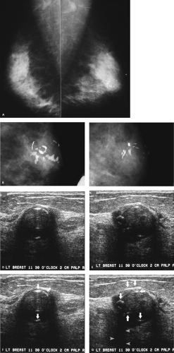
Lymph nodes are present in both axillary regions. A 2-cm mass with obscured margins and associated coarse calcifications is seen in the left subareolar area on the mediolateral oblique views. On the spot compression view, the margins are irregular and indistinct. Compared with the study from 5 years previously, the mass is now more dense and the calcifications are larger.
On physical examination, a readily mobile, hard, nontender mass is palpated at the 11:30 o’clock position, 2 cm from the left nipple. On ultrasound, a round mass with heterogeneous echotexture and shadowing is imaged corresponding to the palpable finding. Several areas of hyperechogenicity (Fig. 1.12F, G, arrows), some curvilinear, others with associated shadowing (Fig. 1.12F, G, arrowheads), are noted consistent with the calcifications seen mammographically. The mammographic findings are diagnostic of a hyalinizing fibroadenoma with associated dystrophic calcifications. Although an ultrasound is shown here for completeness, the diagnosis is made on the mammographic findings. This is explained to the patient; she is reassured that what she feels is a benign lesion that will not turn into cancer and has been present for 5 years with no significant changes. A definitive report is issued describing the palpable finding as a hyalinizing fibroadenoma requiring no further intervention or short-interval follow-up.
As the estrogen levels decrease, the epithelial elements in fibroadenomas atrophy and are replaced by an increasing amount of fibrous tissue. Mammographically, hyalinizing fibroadenomas may decrease slightly in size and become more dense so that, in some women, they become more readily apparent while some undergo calcification, as demonstrated in this patient.
BI-RADS® category 2: benign finding. Next screening mammogram is recommended in 1 year.
Patient 13
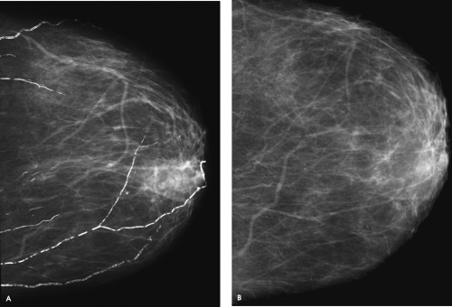 Figure 1.13. Screening mammogram, 63-year-old woman. Craniocaudal (A) view, left breast. Craniocaudal (B) view, left breast, 3 years previously. |
How would you describe the findings?
Linear, parallel, tram-track-like calcifications represent vascular (arterial) calcifications. This patient has developed dense vascular calcifications in the span of 3 years, suggesting the possibility of diabetes or atherosclerotic disease that may be significant. In this patient, I describe the development of vascular calcifications in the mammographic report. It is reasonable to contact the referring physician directly, particularly if the patient provides no information relative to underlying diabetic, cardiac, or renal disease (e.g., what medications is the patient taking?), to discuss the findings.
Stay updated, free articles. Join our Telegram channel

Full access? Get Clinical Tree


