KEY FACTS
Terminology
- •
Fibroid, leiomyoma
- •
Definition: Benign smooth muscle tumor of uterus
Imaging
- •
Typically well-defined round or oval hypoechoic mass ± calcification
- •
Myomas can grow during first 1/2 of pregnancy
- •
Myomas can degenerate during pregnancy
- ○
Most common: Asymptomatic hyaline degeneration
- –
Typically heterogeneous, septated, cystic myoma
- –
- ○
Less common: Symptomatic hemorrhagic degeneration
- –
Hyperechoic, heterogeneous, solid-appearing myoma
- –
Can lead to preterm labor and pregnancy loss
- –
- ○
- •
Placenta can implant upon myoma
- •
Color Doppler findings
- ○
Uterine vessels supply myoma
- –
May see vascular pedicle to pedunculated myoma
- –
- ○
Peripheral swirled pattern of flow is typical
- ○
Clinical Issues
- •
Complications related to size, number, and location
- ○
Lower uterine myoma with ↑ rates of malpresentation, cesarean delivery, and postpartum hemorrhage
- ○
Retroplacental myoma with ↑ rates of placental insufficiency, abruption, and preterm labor
- ○
Multiple myomas associated with fetal growth restriction and postpartum hemorrhage
- ○
- •
Acute pain, low-grade fever associated with hemorrhagic degeneration: MR best to show hemorrhage
Scanning Tips
- •
Note location, type, and size of myoma in every case
- •
Note relationship with placenta and cervix
- •
Adnexal and pedunculated myoma can mimic ovarian mass
- ○
Look for blood supply from uterus
- ○
Look for separate ovary
- ○
- •
If myoma is painful, consider hemorrhagic degeneration and alert provider
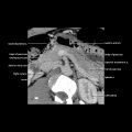
 implants upon a submucosal myoma
  . The myoma is homogeneous and hypoechoic. Placental implantation upon myoma is associated with abruption and fetal growth restriction.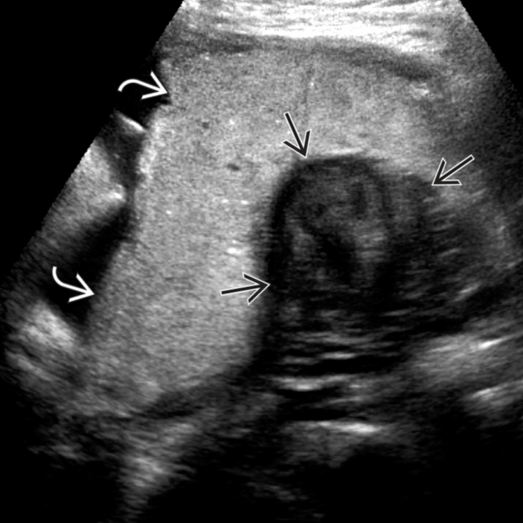
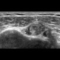
 is implanted upon a myoma
  , demonstrating typical swirled flow pattern on color Doppler. This is probably why a marginal abruption
  occurred in this pregnancy.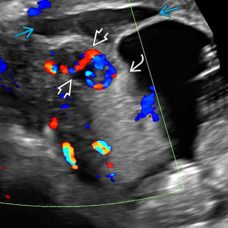
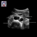
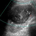
 , the avascular central cystic region and wall nodularities
  mimic an ovarian neoplasm. However, the myoma blood supply is shown coming from the uterus
  and a separate normal ovary was documented (not shown).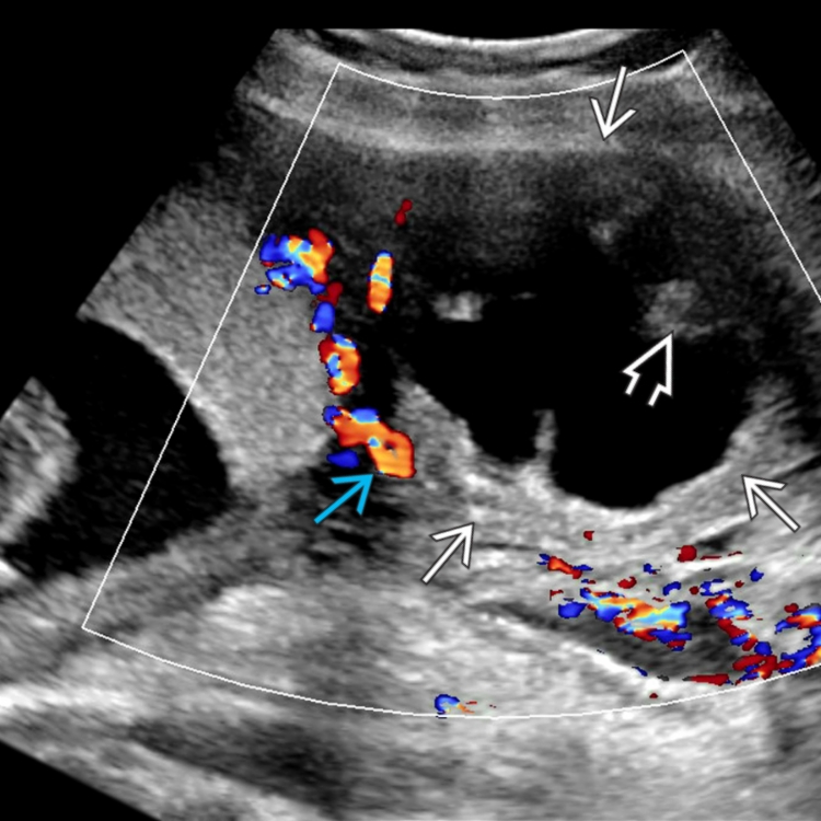
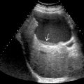
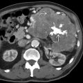
 . Note the internal septations and wall nodularity
  . Smaller uncomplicated solid fibroids
  are also seen.
Related posts:

Stay updated, free articles. Join our Telegram channel

Full access? Get Clinical Tree


