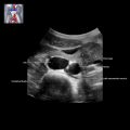
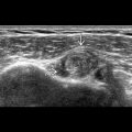
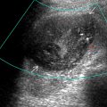
KEY FACTS
Terminology
- •
Mucus-filled cystic dilatation of endocervical gland
- •
Tunnel cluster: Type of nabothian cyst characterized by complex multicystic dilatation of endocervical glands
Imaging
- •
Circumscribed, superficial, simple unilocular cyst in cervix
- ○
Imperceptible thin wall
- ○
Posterior acoustic enhancement
- ○
- •
Average size: 10-15 mm, rarely > 4 cm
- •
Often multiple
- •
May distort endocervical canal or enlarge cervix
- •
Can have proteinaceous debris producing low-level echoes
- •
Rare septations
- •
Can mimic multicystic mass when multiple
- •
Absence of internal or peripheral flow on color Doppler
Top Differential Diagnoses
- •
Adenoma malignum (minimal deviation adenocarcinoma)
- •
Endocervical hyperplasia
- •
Uterine cervicitis
- •
Cystic endometrial polyp
- •
Abortion in progress
- •
Cervical ectopic pregnancy
- •
Cervical cancer
Pathology
- •
Most common pathologic process of cervix: Healing process of chronic cervicitis
Clinical Issues
- •
Incidental finding on imaging or examination, rarely symptomatic from infection or mass effect
- •
More common in women who have had children
- •
Treatment is rarely necessary
Diagnostic Checklist
- •
Requires further evaluation with MR if
- ○
Solid components
- ○
Copious vaginal discharge
- ○
Deep cervical invasion
- ○
Scanning Tips
- •
Use color Doppler if cyst appears potentially solid
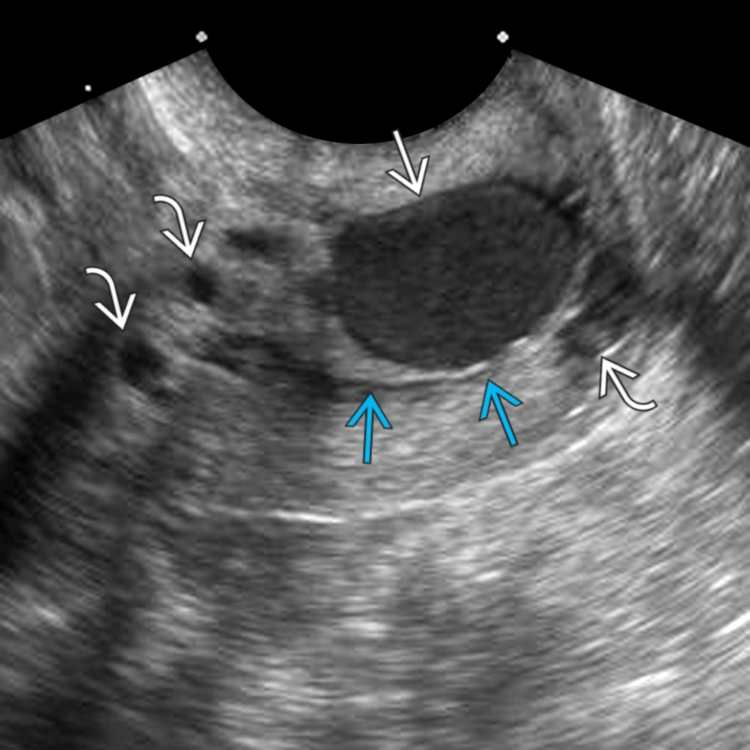
 with posterior acoustic enhancement
  , separate from the endocervical canal
  .
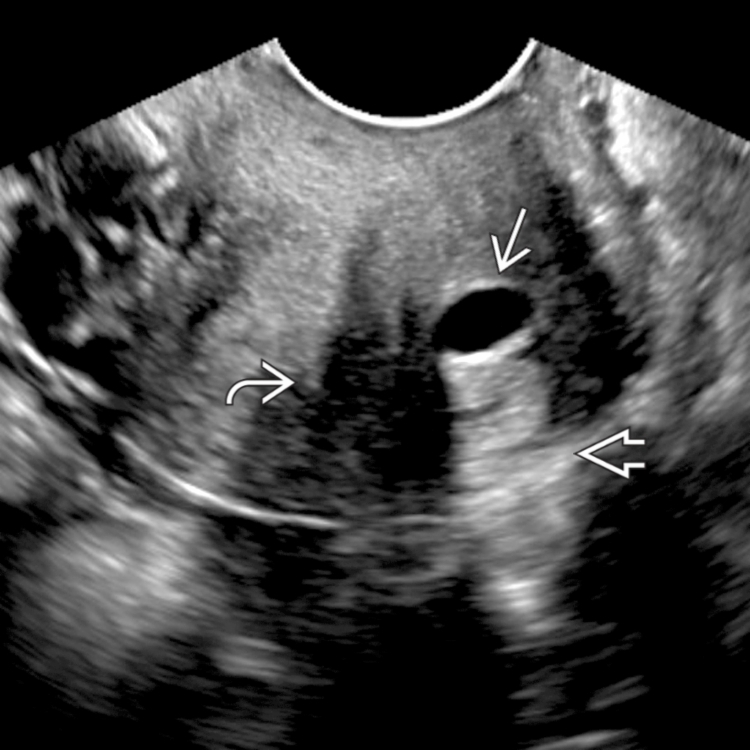
.
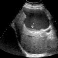
 at the lateral margin of the cervix. Real-time imaging helped confirm the location within the cervical stroma; however, this image demonstrates the potential for misdiagnosis as an ovarian follicle.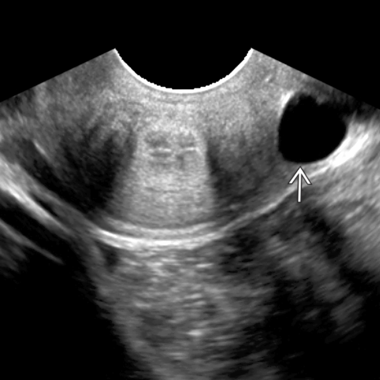
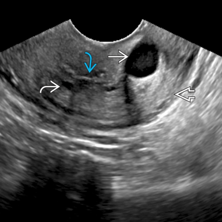
 with internal debris. There is posterior acoustic enhancement
  . Tiny nabothian cysts
  are noted near the endocervical canal
  .
.
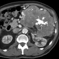
 with homogeneous internal echoes, in addition to multiple smaller nabothian cysts
  . The cervical canal is minimally displaced but remains visible
  .
.