Necrotizing Fasciitis—Pediatric
Andy K. Chon
Daniel B. Nissman
CLINICAL HISTORY
10-month-old boy with left lower extremity erythema and tenderness.
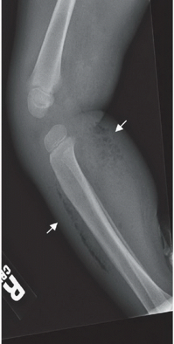 FIGURE 72A |
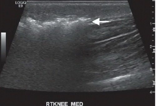 FIGURE 72B |
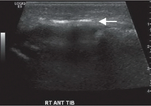 FIGURE 72C |
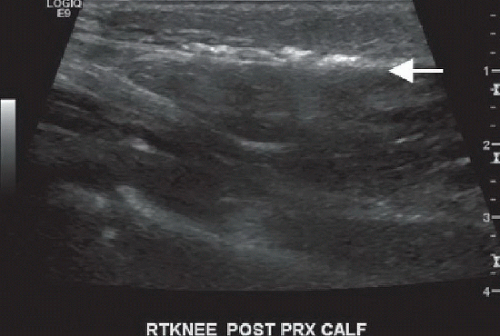 FIGURE 72D |
FINDINGS
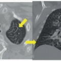
Lateral radiograph of the right leg (Fig. 72A) demonstrates subcutaneous gas along the anterior and posterior lower leg (arrows); the anterior soft tissue gas is oriented in a linear fashion along the interface between the subcutaneous tissues and the underlying muscular compartment, which is invested by the deep fascia. Figures 72B,72C and 72D demonstrate different sonographic appearances of soft tissue gas (arrow), from very echogenic reflectors with extensive posterior streak artifact resulting in complete obscuration of the tissues deep to the gas (Fig. 72B) to much more subtle foci of hyperintensity
along the superficial fascia with relatively preserved appearance of the soft tissues deep to the gas (Fig. 72D).
along the superficial fascia with relatively preserved appearance of the soft tissues deep to the gas (Fig. 72D).
DIFFERENTIAL DIAGNOSIS
Postsurgical changes, recent instrumentation or laparoscopic insufflation, infection with a gas-forming organism (necrotizing fasciitis).
Related posts:


Stay updated, free articles. Join our Telegram channel

Full access? Get Clinical Tree


