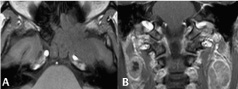
54 Neoplasia I Squamous cell carcinomas are the most common head and neck cancer, arising from the sphenoid sinus in Figs. 54.1A,B. This lesion extends to the nasal cavity and middle cranial fossa, demonstrating a heterogeneous appearance on (A) GRE T1WI. On GRE T1WI, arterial structures demonstrate high SI, highlighting mild compression of the left internal carotid in this instance. (B) Coronal contrast-enhanced T1WI reveals enlarged (short axis >1 cm) enhancing bilateral lymph nodes suspicious for metastatic involvement. Low central nodal SI is consistent with necrosis—a finding suggestive of neoplastic involvement regardless of size. Other malignant features include lack of hilar fat SI, rounded appearance, marginal blurring, infiltration of surrounding fat, or low ADC values. Benign mass-like lesions of the nasopharynx and paranasal sinuses expand rather than invade bone and do not enhance diffusely. An expansile ethmoid mucocele is illustrated in Figs. 54.2A,B, demonstrating fluid-like SI on (A) T2WI and (B) CE T1WI. (B) Faint peripheral enhancement correlates with active mucoperiosteum. If thicker, such enhancement signifies a mucopyocele. Allergic fungal sinusitis and sinonasal polyposis both exhibit similar MRI appearances, but typically involve multiple sinuses. The latter is of inflammatory rather than atopic etiology and is associated with polyps, although these may be seen with allergic fungal sinusitis as well. Such polyps enhance only peripherally.
Related posts:


Stay updated, free articles. Join our Telegram channel

Full access? Get Clinical Tree


