Diagnostic tools
Overactive bladder
Dysfunctional voiding
Medical history
Urgency
Holding maneuvers to contrast urgency
± Increased void frequencya
± Urge incontinence
± Urinary tract infection
± Constipation
Holding maneuvers to postpone micturition
Increased/decreased void frequencya
± Intermittent incontinence
± Urinary tract infection
± Constipation
Bladder diary
Episodes of urgency
Increased number of voidinga
Low voided volumeb
Episodes of incontinence + urge
Amount of urine loss
Bowel movements
Abnormal number of voidinga
Normal/high voided volumeb
Episodes of incontinence without urge
Amount of urine loss
Bowel movements
Uroflowmetry
Tower-shaped curve
Staccato or interrupted curve
Low voided volumeb
Normal/high voided volumeb
Pelvic floor EMG
Relaxed pelvic floor
Overactive pelvic floor
Pre-voiding US
Low bladder capacityb
Normal/increased bladder capacityb
Bladder wall: normal or thickened
Bladder wall: normal or thickened
Postvoiding US
No postvoiding residuum
± Postvoiding residuum
Bladder Diary: Bladder diary records fluid intake, micturition frequency, voided volume, episode of urgency, and urine loss. For diagnostic purpose, it should cover at least 3 days of registration. A bowel movement chart and Bristol stool chart [12] are also important, either separate from or in conjunction with bladder diary.
Urinalysis: It may provide information about urinary tract infection, possible associated diabetes and renal damage, or disease-causing proteinuria. Dipstick may be useful in clinical practice to achieve these information quickly [15].
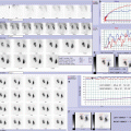
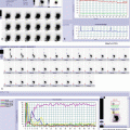
Uroflowmetry (FLW) [5]: It is the less invasive of all urodynamic studies and, therefore, is perfect for pediatric patients [11]. In children, more than one FLW recording should be obtained in the same session before drawing conclusions, and three evaluations are advised [3, 16]. Pelvic floor EMG and ultrasound PVR are recommended, increasing the value of FLW measurement. Results of FLW, combined with those of history, bladder diary, and PVR, seem to be more effective than UDS in detecting both OAB and DV. The shape of the flow curve is the most important factor to analyze in pediatric patients (Fig. 9.1) [5]. In normal voiding, the curve is bell-shaped. OAB may produce an explosive voiding contraction that appears as a high-amplitude flow curve of short duration, namely, a tower–shaped curve. In DV, sphincter overactivity during voiding produces a staccato flow, as a continuous urine flow with periodic reductions in flow rate precipitated by bursts of pelvic floor activity; voiding is commonly prolonged and incomplete. Interrupted or fractionated flow is micturition in separate fractions due to unsustained voiding contractions. Interrupted or fractionated flow can be seen in children with severe DV and UB. Functional and anatomical obstruction often has a low amplitude, and rather even flow curve, that is, a plateau curve. Generally, in children with OAB, voided volume (Vvol) is lower than the expected bladder capacity for age (EBC). On the contrary, in children with DV, Vvol can be higher than EBC. EBC for age is estimated by the formula: [30 + (age in years × 30)] in ml [16]. Evaluating FLW, EBC is of great importance, because it has been shown that uroflow curve changes when the voided volume is less than 50 % of EBC [5]. In children, poor correlation exists between Qmax and outflow resistance. Therefore, Qmax is of minor importance than in adult age.

Fig. 9.1
Uroflowmetry pattern in children with nonneurogenic LUTD. (a) bell-shaped: normal flowmetric curve; (b) Staccato flow indicates dysfunctional voiding; (c) interrupted flow due to severe dysfunctional voiding or underactive bladder

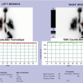
Fig. 9.2
Flowmetry and radiological findings of a 6-year-old girl with dysfunctional voiding, recurrent UTI, and VUR: (a) flowmetric pattern (interrupted) of severe dysfunctional voiding; (b) mild dilation of the right kidney at ultrasound evaluation of the urinary tract; (c) bilateral VUR during voiding at micturition cystourethrography; (d) appearance of VUR at MAG3 cystoscintigraphy
Ultrasound: In all children with proven LUTS, bladder ultrasound is indicated. It should be performed before and after voiding. Prevoid examination contributes to overall assessment of bladder capacity, bladder wall, lower ureteral dilatation, and bladder neck appearance. Since bladder wall thickness depends on the degree of bladder filling and age of children, it is of great variability in pediatric age, and normal values are not disposable. However, correlations have been demonstrated in children with LUTD between bladder wall thickness, urodynamic pattern, and treatment outcome, with good specificity [18]. Therefore, a thickened bladder wall may alert clinicians on the presence of a long-standing LUTD, leading to detrusor hypertrophy. Ultrasound evaluation of bladder immediately after voiding can demonstrate PVR. A PVR ≥10 % of EBC or ≥20 ml is considered significant [5].Related posts:

Stay updated, free articles. Join our Telegram channel

Full access? Get Clinical Tree