, Lawrence A. Zumo2 and Valerie Sim3
(1)
Parkinson’s Clinic of Eastern Toronto, Toronto, ON, Canada
(2)
Silver Spring, Cheverly, MD, USA
(3)
Centre for Prions and Protein Folding Diseases, University of Alberta, Edmonton, AB, Canada
Abstract
A number of degenerative conditions can affect the central nervous system and imaging modalities can be helpful in determining the diagnosis. Here we present cases of frontotemporal dementia, olivopontocerebellar atrophy, spinocerebellar ataxia, multiple systems atrophy, corticobasal degeneration, spinal muscular atrophy and Creutzfeldt-Jakob disease (CJD). CJD is sometimes classified under infectious diseases as it is transmissible, however it is truly a protein folding disease with more similarity to Alzheimer’s and Parkinson’s diseases than any infectious disease, so it is presented here instead.
Case 5.1 Frontotemporal Dementia
An 86 year old male presented with 2 years of gradual onset change in behaviour, lack of interest in activities and memory loss. On examination, frontal release signs were present and he was easily distracted. A CT scan of the brain was performed (Fig. 5.1).
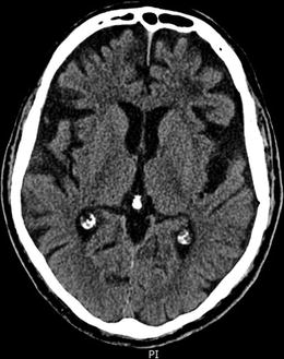

Figure 5.1
Axial CT showing significant bilateral frontal and temporal lobe atrophy and remote left frontal lobe subcortical ischemia
Explanation and Diagnosis
Figure 5.1 shows prominent atrophy of frontal and temporal lobes with relative preservation of the occipital lobes. There is also some remote ischemic change of the left more than right subcortical frontal lobes, consistent with small vessel ischemic damage. This patient has frontotemporal dementia.
Case 5.2 Olivopontocerebellar Atrophy
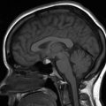
A 37 year old right-handed female presented with speech problems and mild cognitive difficulties, as well as balance problems. Upon physical examination, she had mild dysarthria with scanning speech, finger-nose-finger and heel-knee-shin dysmetria. An MRI of the brain was performed (Fig. 5.2).
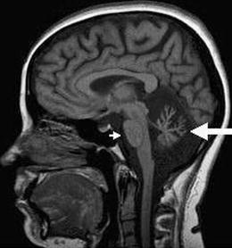

Figure 5.2
Sagittal T1-weighted MRI showing marked pontine (short arrow) and cerebellar (large arrow) atrophy
Explanation and Diagnosis
Figure 5.2 shows striking loss of cerebellar and pontine volume, consistent with a diagnosis of olivopontocerebellar atrophy.
< div class='tao-gold-member'>
Only gold members can continue reading. Log In or Register to continue
Stay updated, free articles. Join our Telegram channel

Full access? Get Clinical Tree


