3 Neuromuscular Diseases
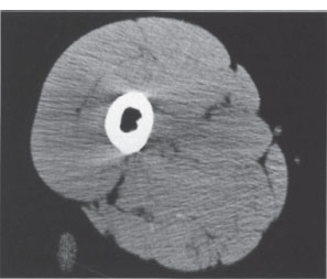
Fig. 3.1 Normal transverse anatomy of the proximal thigh on axial CT. the different soft-tissue structures are distinguished less clearly than on Mr images (Fig. 3.2). Note the artifacts arising from the femur.
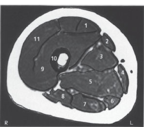
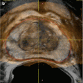
Fig. 3.2 Normal transverse anatomy of the proximal thigh on MRI. T1-weighted SE images provide good delineation of the fascial planes and muscle compartments owing to the high signal intensity of the intermuscular fat.
1 Rectus femoris muscle
2 Sartorius muscle
3 Adductor longus muscle
4 Gracilis muscle
5 Adductor magnus muscle
6 Semimembranosus muscle
7 Semitendinosus muscle
8 Biceps femoris muscle (long head)
9 Vastus intermedius muscle
10 Femur
11 Vastus lateralis muscle
Imaging goals in neuromuscular diseases
 Detect pathologic changes
 Determine the location and extent of such changes
 Locate a suitable biopsy site
 Differentiate true muscular hypertrophy from pseudohypertrophy
Neuromuscular imaging, then, requires procedures that offer the highest possible spatial and contrast resolution. While plain radiographs can essentially detect only bony changes and softtissue calcifications, sonography and especially computed tomography (CT) and magnetic resonance imaging (MRI) can also discriminate muscle tissue, connective tissue, and fatty tissue and detect abnormalities of these structures. Myosonography has notyet become an established modality because of its poor spatial resolution and its inability to define deeply situated muscles.
CT offers better spatial resolution, and its sectional capabilities are particularly useful for diagnosing diseases of the limb muscles, neck muscles, and the muscles of the shoulder girdle and pelvic girdle (Fig. 3.1).
Normal skeletal muscles have an attenuation value of 30–90 Hounsfield units (HU). The fasciae are more attenuating than muscle, but they are so thin that usually they are visible only around larger muscle groups. Because of the large attenuation difference between bone and soft tissues, streak-like beam-hardening artifacts are common and can lead to errors of visual interpretation and density measurement.
MRI also has high spatial resolution but is distinguished by its superior soft-tissue contrast, its multiplanar capabilities, and its ability to provide largely artifact-free images. The dominant technique is spin-echo (SE) imaging, which can discriminate cortical bone (signal void), connective tissue (hypointense), fat (hyperintense), and muscle tissue (intermediate) based on the different relaxation times of the different tissues (Fig. 3.2).
Because the various muscles and muscle groups are bounded by fat-containing septa, they are easy to identify on magnetic resonance (MR) images.
MR spectroscopy (MRS) can also yield important diagnostic information. 31
Stay updated, free articles. Join our Telegram channel

Full access? Get Clinical Tree


