Chapter 14 New technology and recent advances in ultrasound imaging
DIGITAL BEAM FORMING
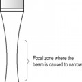
Using analog beam formers, it is not possible to produce delay timers with the accuracy required and this limits their performance. However, beam formers are now produced using digital electronics for the time delays. In addition to producing narrower beams, digital beam formers can operate at higher frequencies and can be used with broad-bandwidth transducers.
EXTENDED FIELD OF VIEW IMAGING
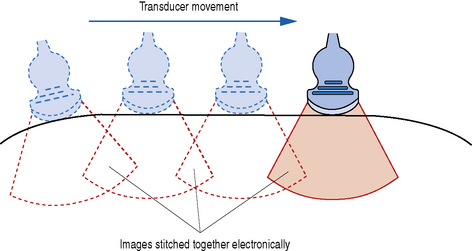
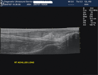
This is an imaging process which combines static B-mode techniques with real-time imaging so that a large subject area can be viewed on a single static image. Extended field of view (FOV) images are obtained by sliding the probe over the area of interest and as the images are acquired they are ‘stitched together’ electronically (Fig. 14.1). The result is a single slice image covering the whole area of interest, for example a full length view of the Achilles tendon (Fig. 14.2). Image feature recognition software is used to combine images.
COMPOUND IMAGING
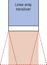
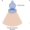
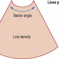
This technique combines electronic beam steering with conventional linear array technology to produce real-time images acquired from different view angles (see Fig. 14.3) Between 3 and 9 sector images are rapidly acquired and combined to produce a compound real-time image (see Fig. 14.4). Compound imaging improves image quality by reducing speckle, clutter, and other acoustic artifacts. It also gives better definition of the boundaries of structures. Because of the improved contrast resolution, compound imaging may be useful for the breast, peripheral blood vessels, and musculoskeletal applications.
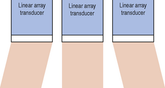
Fig. 14.3 Diagram showing how electronic beam steering is used to acquire images from different angles
THREE-DIMENSIONAL IMAGING
3-D Imaging Technology
The production of a 3-D image requires a volume of tissue to be scanned. The data from this volume are then used to construct the types of image required. There are three approaches to scanning a volume of tissue: free-hand, mechanical, and electronic scanning.
Free-hand 3-D imaging
In this approach the operator sweeps the probe across the volume of interest and a series of scanning planes are recorded according to their position on the patient (see Fig. 14.5). In order to register these planes, a method of determining the position of the transducer in space is required. This can be achieved by using a receiver in the probe which will detect a magnetic field generated by a transmitter situated next to the couch (see Fig 14.6). Each image slice will have image information and position information for use in 3-D construction. Another method of determining the position of the scan plane is to use a radio transmitter and radio detection coils attached to the probe.

Stay updated, free articles. Join our Telegram channel

Full access? Get Clinical Tree


