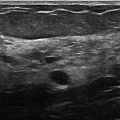
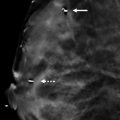
Presentation and Presenting Images
( ▶ Fig. 7.1, ▶ Fig. 7.2, ▶ Fig. 7.3, ▶ Fig. 7.4, ▶ Fig. 7.5, ▶ Fig. 7.6, ▶ Fig. 7.7, ▶ Fig. 7.8, ▶ Fig. 7.9)
A 43-year-old female presents for routine screening mammography.
7.2 Key Images
( ▶ Fig. 7.10, ▶ Fig. 7.11, ▶ Fig. 7.12, ▶ Fig. 7.13)
7.2.1 Breast Tissue Density
There are scattered areas of fibroglandular density.
7.2.2 Imaging Findings
Conventional mammography ( ▶ Fig. 7.1, ▶ Fig. 7.2, ▶ Fig. 7.3, and ▶ Fig. 7.4), synthetic mammography ( ▶ Fig. 7.5, ▶ Fig. 7.6, ▶ Fig. 7.7, and ▶ Fig. 7.8), and tomosynthesis imaging (not shown) demonstrate no suspicious calcifications, asymmetries, masses, or architectural distortions in either breast.
The inferior aspect of the right breast on the mediolateral oblique (MLO) view is incompletely imaged ( ▶ Fig. 7.3). A repeat right MLO view is shown in ▶ Fig. 7.9.
The skin line is absent from both the synthetic mammograms (arrow in ▶ Fig. 7.10 and ▶ Fig. 7.12) and tomosynthesis imaging (not shown). The skin line is clearly seen on the conventional mammograms (arrow in ▶ Fig. 7.11 and ▶ Fig. 7.13).
7.3 BI-RADS Classification and Action
Category 1: Negative
7.4 Differential Diagnosis
Technical artifact: There was a detector failure on the tomosynthesis movie, which resulted in nonvisualization of the skin edge on both the digital breast tomosynthesis (DBT) movies and the synthetic mammogram. This did not affect the conventional digital mammogram.
Negative/benign study: There are no suspicious findings in either breast; however, this case is not “completely negative” due to positioning.
Missed finding: Sometimes mammograms are interpreted as negative or benign when there is an abnormality. There is not a missed finding on this mammogram.
7.5 Essential Facts
The imaging from this case was reviewed by imaging physicists and the loss of the skin line was believed secondary to undercompression of the breast tissue during the examination. The compression thickness for the current study was 80 mm compared to 60 mm on the prior study.
Artifacts can be subtle and escape detection. Tomosynthesis is a relatively new modality, and, with each new modality, there is a learning curve for both technologists and radiologists.
Each view in this case for the conventional mammography, synthetic mammography and tomosynthesis studies was performed without repositioning the patient. This is why the right MLO view is poorly positioned on each of the three studies. A conventional right MLO view was repeated later.
Positioning and obtaining adequate compression are important for both conventional mammography and tomosynthesis.
7.6 Management and Digital Breast Tomosynthesis Principles
High-quality mammograms should be performed at the lowest radiation dose possible.
Mammographic compression is important because it immobilizes the breast, reducing motion and geometric blur, and it prevents overpenetration by the radiation beam by decreasing the thickness of the breast.
The Monte Carlo study of mass and microcalcification conspicuity in tomosynthesis1 showed that reduced compression will have a minimal effect on the radiologist’s performance interpreting tomosynthesis studies.
Less compression will increase women’s comfort with DBT; however, adequate compression must be applied to prevent artifacts.
Technologists and radiologists must be aware of artifacts associated with DBT. As we gain more experience, we will easily detect and correct the artifacts.
7.7 Further Reading
[1] Saunders RSJr, Samei E, Lo JY, Baker JA. Can compression be reduced for breast tomosynthesis? Monte Carlo study on mass and microcalcification conspicuity in tomosynthesis. Radiology. 2009; 251(3): 673‐682 PubMed

Fig. 7.1 Right craniocaudal (RCC) mammogram.
Stay updated, free articles. Join our Telegram channel

Full access? Get Clinical Tree


