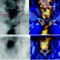
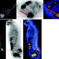
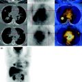
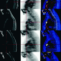
Fig. 11.1
The PET demonstrates the high carbohydrate metabolism of both masses (arrows)
No other focal lesions. Histological re-evaluation needed.
CT: massive submandibular solid, patchy lymph node swelling with irregular margins, inseparable from the adjacent tissues. It is associated in the ipsilateral deep lateral cervical region with adenopathy with similar densitometric characteristics.
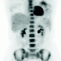
The MIP image:
allows you to appreciate the extent of the massive lymph node involvement and the intensity of FDG consumption;
excluded abnormal metabolism of the mediastinal mass on CT reported in medical history;
Stay updated, free articles. Join our Telegram channel

Full access? Get Clinical Tree


