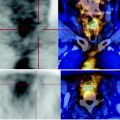
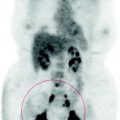
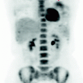
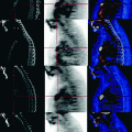
Fig. 35.1
MIP Image: mediastinal lymphadenopathy with elevated glucose metabolism
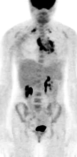
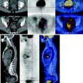
Fig. 35.2
MIP Image: mediastinal lymphadenopathy with elevated glucose metabolism
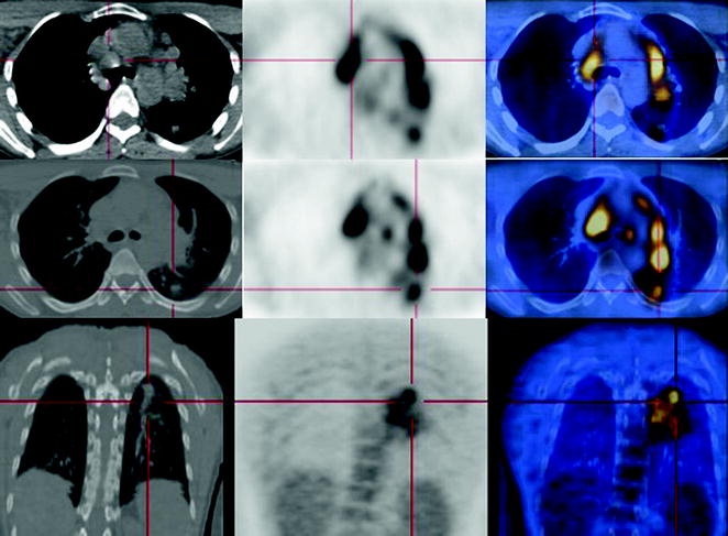
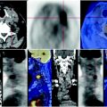
Fig. 35.3


PET-CT in the axial sections shows diffuse involvement of mediastinal lymph nodes, characterized by a high metabolism. At the posterior segment of the upper lobe of the left lung, the coronal reconstruction documents an ill-defined, inhomogeneous coarse parenchymal consolidation, with irregular margins, which has low metabolic activation resulting from inflammatory reactive rearrangement, probably due to embolism. A primary pulmonary neoplasm is unlikely
Stay updated, free articles. Join our Telegram channel

Full access? Get Clinical Tree


