• An incompetent mitral valve allowing regurgitant flow from the left ventricle (LV) into the left atrium (LA) during systole • A very large left atrium is more commonly seen with long-standing mitral regurgitation than with mitral stenosis • Mitral annulus calcification: this rarely occurs before 70 years of age (F>M) • Dystrophic calcification of the mitral valve: unlike mitral annulus calcification, this is very suggestive of a rheumatic aetiology • This is usually due to chronic rheumatic fever resulting in a mixture of stenosis and regurgitation (but both cannot be severe at the same time) • The resultant increased left atrial pressure leads first to interstitial and then alveolar pulmonary oedema • Left atrial enlargement: the left atrial appendage is particularly affected • Parenchymal lung changes of haemosiderosis and intrapulmonary ossification: these may appear after several years of pulmonary venous congestion • Curvilinear calcification: this may occur within the left atrial wall or within the clot lining the wall • Congenital mitral stenosis is rare • Long-standing mitral stenosis can result in atrial fibrillation (complicated by left atrial thrombus) Causes of mitral regurgitation • Acute aortic regurgitation: causes include bacterial endocarditis or (rarely) occurring after trauma or aortic dissection • Chronic aortic regurgitation: congenital deformities (e.g. bicuspid aortic valve or Marfan’s syndrome) • Acute disease: ventricular compliance cannot compensate • Chronic disease: ventricular compliance compensates • Calcific aortic stenosis: most commonly due to degenerative calcium deposition on normal aortic cusps (cf. mitral stenosis with calcium is deposition on a stenosed valve) • Previously commonly due to calcification of a congenitally deformed bicuspid valve • Rheumatic aortic stenosis: inflammatory fusion of aortic valve cusp commissures Calcified valves appear as a signal void • It can demonstrate impaired aortic valve opening (and degree of stenosis), valve morphology and left ventricular function (± hypertrophy) • Systolic flow dephasing within the aortic root has a loose relationship to the severity of the stenosis • Diastolic flow dephasing within the left ventricular outflow tract can assess any associated aortic regurgitation • Non-specific cardiac enlargement • In rheumatic heart disease the features of mitral stenosis predominate (left atrial enlargement and pulmonary arterial enlargement) • Tricuspid valve calcification may be seen (dystrophic degeneration from ageing as well as chronic severe right ventricular hypertension)
Non-ischaemic acquired heart disease
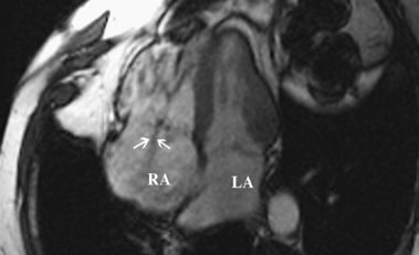
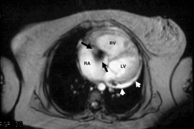
ACQUIRED MITRAL VALVE DISEASE
MITRAL REGURGITATION
DEFINITION
PEARLS
 it is seen with hypercalcaemic states (e.g. end-stage renal disease)
  it may lead to mild mitral regurgitation (but rarely stenosis)
  there is an increased risk of infective endocarditis
  it is also associated with transient ischaemic attacks (due to emboli or a carotid stenosis) and atrioventricular conduction disturbances
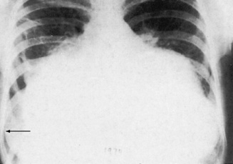
MITRAL STENOSIS
DEFINITION
 secondary pulmonary arterial hypertension may develop, leading to pulmonary valve regurgitation, right ventricular dilatation and functional tricuspid regurgitation (there may also be tricuspid valve rheumatic involvement with additional stenosis or regurgitation)
RADIOLOGICAL FEATURES
CXR
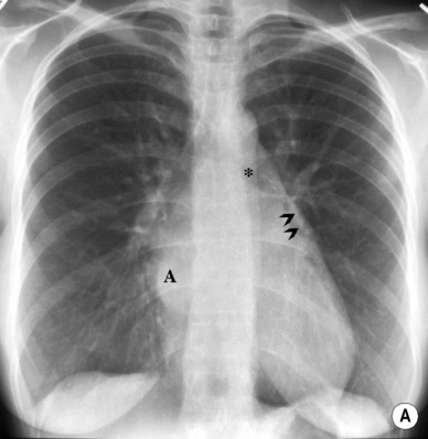
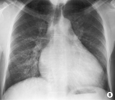
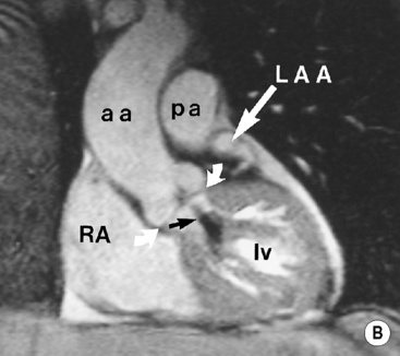
 appearances can vary from a simple straightening of the left heart border to a large bulge at the site of the appendage
  a grossly dilated left atrium can enlarge to the right and also posteriorly (causing oesophageal displacement and dysphagia)
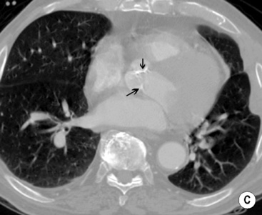
 A ‘double density’ behind the heart
 Widening of the subcarinal angle
 Left ventricular enlargement is not a feature (cf. mitral regurgitation)
PEARLS
Valve abnormalities
Supporting structure abnormalities
Acute rheumatic mitral valve disease
Chordal rupture (e.g. post MI)
Mitral valve prolapse
Papillary muscle rupture dysfunction
Bacterial endocarditis
Functional mitral regurgitation
Prosthetic valve leaks
Mitral annular calcification
Connective tissue diseases (e.g. SLE/RA)
Atrial myxoma
ACQUIRED AORTIC VALVE DISEASE
AORTIC REGURGITATION
Definition
 it develops rapidly with increasing left ventricular end-diastolic pressure and acute heart failure
 rheumatic heart disease
  syphilitic aortitis
  ankylosing spondylitis
  a descending aortic aneurysm
Chronic
Pearls
 it is associated with a very large rise in ventricular end-diastolic pressures (limiting regurgitant flow)
 end-diastolic pressures remain low and the patient remains asymptomatic until heart failure develops (with a worsening prognosis)
AORTIC STENOSIS
Definition
 often associated with aortic regurgitation + mitral valve involvement
  associated mitral valve disease can cause severe dyspnoea
Radiological features
TRICUSPID AND PULMONARY VALVE DISEASE
TRICUSPID VALVE DISEASE
TRICUSPID REGURGITATION
TRICUSPID STENOSIS
CXR
 there may be dilatation of the superior and inferior vena cava
PULMONARY VALVE DISEASE
PULMONARY STENOSIS
PULMONARY REGURGITATION

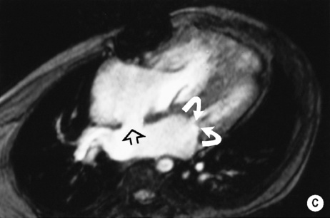
 pulmonary oedema (flow dynamics localizes this to the right upper zone)
 it can assess any regurgitant jet direction (with Doppler assessment)
 phase contrast CMR can be used as a substitute for Doppler in assessing valve area
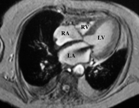
 pulmonary oedema with left heart failure (an important cause of pulmonary oedema with a normal-sized heart)
  a normal aorta (unless there is associated aortic disease causing dilatation – e.g. Marfan’s syndrome)
 moderate thoracic aortic enlargement (severe enlargement with aortitis or chronic dissection)
  infrequent valve calcification
  left atrial enlargement may dominate (if mitral valve disease is present)
 it also allows assessment of LV function
 chest pain
  left ventricular hypertrophy (LVH)
 ultrafast CT can assess the severity of aortic stenosis by imaging the valve opening
 post-stenotic dilatation is rare
  gross valvular calcification is rare
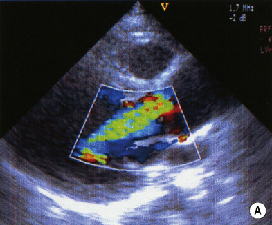
 endocarditis (often as a complication of IV drug abuse)
  pulmonary hypertension (caused by the associated dilated right ventricle)
  previous mitral valve replacement
  Ebstein’s anomaly
  endomyocardial fibrosis
 a low pressure drop across the valve is seen with severe disease
 carcinoid syndrome
  tumours (especially right atrial myxoma)
  endocarditis
 rheumatic heart disease (very rare)
 tumour
 rubella
  tumour
  thrombus
  surgical banding
  Takayasu’s aortoarteritis
  Behcet’s disease
 right ventricular hypertrophy
 pre- and post-stenotic dilatation occurs with dilatation of the right ventricle and central pulmonary arteries