Presentation and Presenting Images
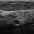
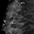
( ▶ Fig. 21.1, ▶ Fig. 21.2, ▶ Fig. 21.3, ▶ Fig. 21.4)
A 60-year-old female presents for asymptomatic screening mammography.
21.2 Key images
( ▶ Fig. 21.5, ▶ Fig. 21.6, ▶ Fig. 21.7, ▶ Fig. 21.8)
21.2.1 Breast Tissue Density
There are scattered areas of fibroglandular density.
21.2.2 Imaging Findings
Within the subareolar region of the right breast, there appears to be a more focal area of fibroglandular tissue compared to the remaining breast tissue ( ▶ Fig. 21.5 and ▶ Fig. 21.6). This is seen on the conventional digital mammograms and the synthetic mammograms. The digital breast tomosynthesis (DBT) images ( ▶ Fig. 21.7 and ▶ Fig. 21.8) reveal that this is part of a mass with circumscribed margins containing fat and fibroglandular components. The left breast is normal (not shown).
21.3 BI-RADS Classification and Action
Category 2: Benign
21.4 Differential Diagnosis
Hamartoma: This mass is present in a fatty to scattered density background. This background can make masses that have a high fat component difficult to identify. The pseudocapsule seen on DBT supports the diagnosis of a hamartoma.
Lipoma : Lipomas are encapsulated fatty masses; however, they do not contain fibroglandular components.
Normal fibroglandular tissue: On conventional mammography, this mass appears to blend into the other tissues in the subareolar region. The DBT helps make clear the capsule, which indicates that this mass is a hamartoma.
21.5 Essential Facts
Hamartomas can be found at any age.
Hamartomas are often referred to as a ‘breast within a breast.’
The mammographic appearance of hamartomas is variable due to the variation in the amount and types of breast tissue elements that they can contain.
Typically hamartomas occur as single entities. It is rare for a patient to have multiple hamartomas (unlike fibroadenomas).
A breast mass that is encapsulated and contains a mixture of adipose and fibroglandular components can be considered benign.
21.6 Management and Digital Breast Tomosynthesis Principles
The superior marginal detail of DBT allows for more accurate analysis of masses.
DBT allows for the recognition of benign encapsulated fat-containing masses, when conventional mammography would have dictated the need for further evaluation.
The ability to scroll through lesions on DBT allows better assessment of margins of masses.
A mass that fulfills the criteria of a hamartoma, encapsulated and containing fatty and fibrous components, does not need further diagnostic evaluation unless there are findings that could represent an associated suspicious finding. DBT will likely aid in less recall mammography of these benign masses due to the ability to better define the margins of the mass with DBT.
21.7 Further Reading
[1] Conant EF. Clinical implementation of digital breast tomosynthesis. Radiol Clin North Am. 2014; 52(3): 499‐518 PubMed
[2] Freer PE, Wang JL, Rafferty EA. Digital breast tomosynthesis in the analysis of fat-containing lesions. Radiographics. 2014; 34(2): 343‐358 PubMed

Fig. 21.1 Right craniocaudal (RCC) mammogram.
Stay updated, free articles. Join our Telegram channel

Full access? Get Clinical Tree


