25 Nonvascular Pathology Encountered During Venous Sonography
Numerous findings may be encountered during the course of a venous examination. These findings may indicate the cause of extremity pain or swelling apart from venous disease. Recognition of the sonographic features associated with these pathologies is critical for accurate diagnosis.1–12 Many of these diagnoses are common, such as edema, hematoma, lymph nodes, or popliteal (Baker’s) cysts. Other findings are less common, but no less important, including abscess, joint effusion, adenopathy, benign or malignant tumor, and metastatic disease.
Soft Tissue Edema
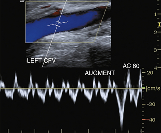
There is a change in venous flow patterns associated with CHF. Normally, phasic, nonpulsatile waveforms are seen in the lower extremity veins. In the setting of CHF, the waveforms are usually more pulsatile and typically bidirectional, with components seen above and below the baseline2 (Figure 25-1). As noted in Chapter 1, causes of increased venous pressure such as CHF or tricuspid insufficiency lead to increased transmission of cardiac phasic changes in pressure and blood flow to the peripheral veins of the upper and lower limbs. These changes result in increased pulsatility in the peripheral vein waveforms. Pulsatility in the venous system may resemble arterial pulsations because of the bidirectional pattern of blood flow. Comparison of the arterial and venous flow patterns is key to distinguishing pulsatile venous signals from arterial flow. On close inspection, one will note that the phases of the venous pattern are less periodic than the typical triphasic waveforms found in the arterial system. Other clues to the nature of the flow abnormality are the direction of flow and the communications with the superficial veins. Changes in venous flow will also be seen with the Valsalva maneuver.
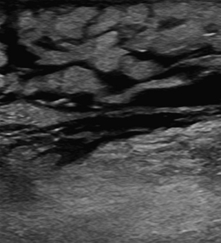
Gray-scale imaging may show a marbled, reticulated pattern when edema collects in the subcutaneous fat. Small collections of fluid may be seen when edema collects in the superficial tissues (Figure 25-2). This can be associated with significant soft tissue swelling and attenuation of sound that can restrict visualization of deep tissues and underlying vascular structures. A “sound” approach to this problem is to switch transducers to allow lower-frequency imaging. Changing from the usual 5- to 7-MHz linear array probe to a 2- to 5-MHz curved array transducer should allow deeper penetration of sound and visualization of the area of interest.
Bilateral leg swelling is a typical presentation for patients with venous congestion. It is also a common indication for venous sonography. Studies have shown that the likelihood of identifying DVT in patients with bilateral leg swelling and no significant risk factors is low (≤5%).3 Naidich and associates4 showed that 23% of patients with bilateral symptoms and risk factors had underlying DVT. In this group, 78% of patients had risk factors for DVT.5 DVT cannot be reliably diagnosed or excluded on clinical grounds alone.
Lymphedema
Lymphedema is also associated with leg swelling and mimics DVT.5 Obstruction of the lymphatics due to malignancy, trauma, or surgery will cause extremity swelling and pain. Patients will typically present with unilateral or bilateral leg swelling. The appearance of lymphedema is indistinguishable from soft tissue edema associated with venous congestion. There may or may not be lymph node enlargement to suggest the etiology of the leg swelling.
Hematoma
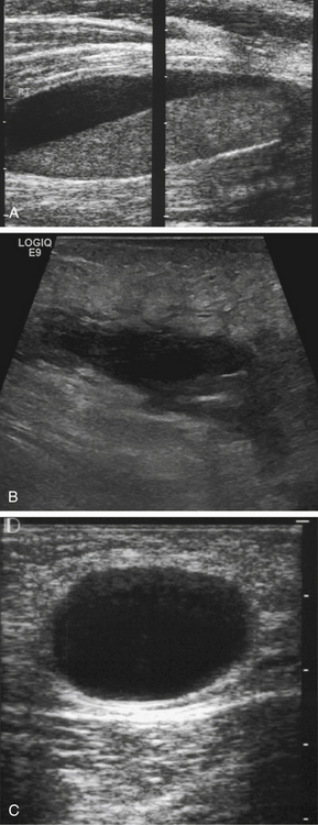
Sonographic examination of a focal mass will reveal a cystic, complex, or solid lesion, which is typically found in the superficial tissues. The mass may initially have irregular borders and demonstrate areas of fluid and solid tissue depending on the amount of clot in the hematoma (Figure 25-3). Over time, the mass will retract and become better defined and ovoid. Hematoma may also be spread through fascial or muscle planes and seen as hypoechoic or isoechoic tissue. This may be very difficult to identify as there is no significant difference in echogenicity from adjacent structures. Leg swelling may be the only clue to the soft tissue infiltration with hematoma. Magnetic resonance imaging (MRI) provides superior soft tissue contrast and displays the extent of soft tissue hematoma.
Related posts:
 Normal Cerebrovascular Anatomy and Collateral Pathways
 Ultrasound Assessment of Lower Extremity Arteries
 Ultrasound Assessment of the Vertebral Arteries
 Arterial Anatomy of the Extremities
 Duplex Ultrasound Evaluation of the Uterus and Ovaries
 Extremity Venous Anatomy and Technique for Ultrasound Examination
Stay updated, free articles. Join our Telegram channel

Full access? Get Clinical Tree


