, Marilyn J. Siegel2, Tomasz Miszalski-Jamka3, 4 and Robert Pelberg1
(1)
The Christ Hospital Heart and Vascular Center of Greater Cincinnati, The Lindner Center for Research and Education, Cincinnati, OH, USA
(2)
Mallinckrodt Institute of Radiology, Washington University School of Medicine, St. Louis, Missouri, USA
(3)
Department of Clinical Radiology and Imaging Diagnostics, 4th Military Hospital, Wrocław, Poland
(4)
Center for Diagnosis Prevention and Telemedicine, John Paul II Hospital, Kraków, Poland
Abstract
There are many not uncommonly seen variants that may be mistaken as abnormal, and recognition of these normal variants will increase diagnostic imaging accuracy and reduce the incidence of false alarms.
There are many not uncommonly seen variants that may be mistaken as abnormal, and recognition of these normal variants will increase diagnostic imaging accuracy and reduce the incidence of false alarms.
3.1 Left Ventricular Diverticula
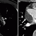
Congenital left ventricular (LV) diverticula (Figs. 3.1 and 3.2) are defined as any outpouching from the ventricle that is lined by ventricular myocardium. Its connection to the ventricle may be wide or narrow [1].
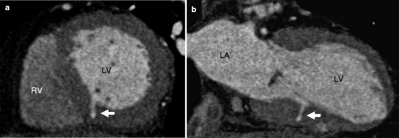
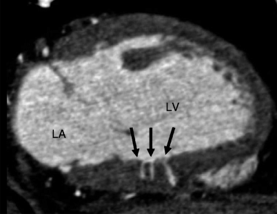

Fig. 3.1
Left ventricular (LV) diverticulum. Panels (a) (short axis) and (b) (long axis) demonstrate a single diverticulum (white arrows) located at mid-ventricle level at the inferior right ventricular insertion site. LV left ventricle, RV right ventricle, LA left atrium (Image generously provided by Jill E. Jacobs, MD, FACR, Professor of Radiology, New York University Langone Medical Center)

Fig. 3.2
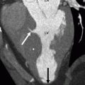
Multiple diverticula are seen in diastole (arrows). Diverticula must be differentiated from contained myocardial rupture which is a highly malignant finding. Contained rupture typically appears as a spiral dissection through necrotic tissue (not at acute angles) and is associated with a focal area of hypoperfusion, wall motion abnormality, and thrombotic occlusion of the associated coronary artery. LV left ventricle, LA left atrium (Image generously provided by Jill E. Jacobs, MD, FACR, Professor of Radiology, New York University Langone Medical Center)
The incidence of congenital ventricular diverticula has been reported to be between 0.04 and 2.2 % in an autopsy series of patients dying of cardiac disease [1–3]. LV diverticula range in size from 0.5 to 9 cm in diameter and have been reported in nearly all LV locations. Most commonly, however, they are found in the left ventricular apex and along the inferior or inferoseptal wall left ventricular walls and at the right ventricular insertion point [4]. When multiple diverticula are present (40 % of the cases, Fig. 3.2), they tend to be in close proximity to each other [1].
The Cantrell syndrome is defined as the association of LV diverticula with congenital midline thoracoabdominal abnormalities, sternal and diaphragmatic defects, and partial absence of the inferoapical pericardium [5].
Congenital ventricular diverticula should be differentiated from acquired causes of ventricular aneurysms and pseudoaneurysms such as those that occur after myocardial infarction, myocardial inflammation, or cardiac trauma. These entities may be similar in appearance making differentiation by noninvasive imaging modalities challenging. Often, the correct diagnosis is made using associated clinical history and imaging abnormalities (h/o trauma or myocardial infarction with associated imaging abnormalities like ventricular thinning or calcification). In addition, acquired ventricular aneurysms consist of predominantly fibrous tissue and are associated with abnormal cardiac contour in diastole and myocardial bulging during systole.
Note that muscular ventricular septal defects that spontaneously close by overlapping ventricular trabeculations may also produce diverticular outpouching of the ventricle.
Ventricular noncompaction may have a similar appearance to that of a ventricular diverticulum. The diagnosis of ventricular noncompaction is confirmed by identifying more than three deep intertrabecular recesses and a systolic ratio of noncompacted to compacted layers of >2.2 [6, 7]. Ventricular noncompaction is discussed in detail in Part 4, Chap. 9.
3.2 Myocardial Bridging
Myocardial bridging (Fig. 3.3) is defined as the presence of an intramyocardial segment of a major coronary artery that normally has an epicardial course. The frequency of myocardial bridging observed by computed tomographic angiography (CTA) is nearly 60 % [8]. Myocardial bridging is typically a benign finding and the vast majority of myocardial bridges are asymptomatic. It is clinically significant only when associated with regional hemodynamic alterations. Hemodynamically significant findings typically occur only when myocardial bridging involves the left anterior descending artery. Even in adult patients with hypertrophic cardiomyopathy, myocardial bridging is not associated with increased risk of death, including sudden cardiac death.
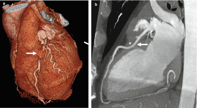

Fig. 3.3
Myocardial bridging. Panel (a) (3D volume-rendered image) shows the “bridge” of myocardium overlying the proximal left anterior descending artery (white arrow). Panel (b) (maximum intensity projection in a transverse plane) illustrates the proximal left anterior descending artery coursing within the myocardium (white arrow) and the overlying “bridge of myocardium” (white asterisk)
Note that myocardial bridging is almost never seen in the right coronary artery since most of the course of the right coronary artery is not near the myocardium.
3.3 Left Ventricular Trabeculations
Prominent left ventricular trabeculations (Fig. 3.4) have been reported in 323 of 474 autopsies (68 %) in normal human hearts and they occur equally in males and females (72 and 65 %, respectively) [9]. No variation is noted when age is considered [9]. Accordingly, prominent left ventricular trabeculations are considered to be common variants of the normal human heart, frequently visualized during CTA examinations, and these should not to be mistaken for isolated left ventricular noncompaction (described above).
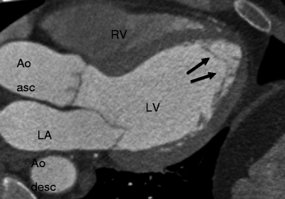

Fig. 3.4
Normal left ventricular trabeculations (arrows) not associated with papillary muscles. These do not meet the criteria for isolated left ventricular noncompaction. LV left ventricle, RV right ventricle, LA left atrium, Ao Asc ascending aorta, Ao Desc descending aorta
3.4 Papillary Muscle Attachment to the Left Ventricular Free Wall
Stay updated, free articles. Join our Telegram channel

Full access? Get Clinical Tree


