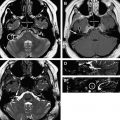
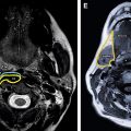
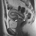
The diagnostic usefulness of abdominal magnetic resonance (MR) imaging lies in the improved contrast resolution and ability to qualify several tissue characteristics of a specific organ or lesion. Our institution uses organ-specific protocols to facilitate technical reproducibility and optimize scan duration. These protocols are discussed individually in this article when applicable, noting that many build on a basic protocol with slight variations. Because most abdominal MR imaging studies are targeted toward an organ or area of interest, this article discusses the protocol strategies and relevant anatomy in a segmented/organ-specific manner.
Abdominal magnetic resonance (MR) imaging has traditionally been used as a problem-solving method after a previous indeterminate imaging investigation such as computed tomography (CT) or ultrasound, frequently using organ-specific protocols to highlight the condition in question. Increasingly, abdominal MR imaging is being used as a primary imaging investigation with expansion of clinical indications and increased awareness of cumulative ionizing radiation dose in medical imaging. Therefore, it is important to understand the normal abdominal anatomy on MR imaging, particularly the visceral organ appearances, as well as commonly encountered variants and disease mimics. Because most abdominal MR imaging studies are targeted toward an organ or area of interest, this article discusses the protocol strategies and relevant anatomy in a segmented/organ-specific manner.
In general, abdominal MR imaging benefits substantially from the use of a high field strength magnet (1.5 Tesla or greater) and local phased-array surface coils around the anatomy of interest. Most commonly, breath-held sequences with high temporal resolution and moderate spatial resolution are frequently used in abdominal MR imaging to mitigate the effects of respiratory and bowel motion. However, when high spatial resolution and improved image signal are required, motion compensation techniques such as respiratory triggering or diaphragm-navigation can be used to facilitate longer acquisition times. The diagnostic usefulness of abdominal MR imaging lies in the improved contrast resolution and ability to qualify several tissue characteristics of a specific organ or lesion. Generally, abdominal MR imaging protocols include precontrast T1-weighted images, T2-weighted images, and dynamic postcontrast T1-weighted images with sufficient imaging redundancy of the organ system in question. Our institution uses organ-specific protocols to facilitate technical reproducibility and optimize scan duration. These protocols are discussed individually in this article when applicable, noting that many build on a basic protocol with slight variations ( Table 1 ).
| Abdominal/Liver Protocol | |||||||
|---|---|---|---|---|---|---|---|
| Coronal T2 | Axial T2 | Axial In/Opposed T1 | Axial Fat- Suppressed T2 | Axial Diffusion | Axial Pre/Post T1 | Axial Dynamic Pre/Post T1 | |
| Pulse sequence | SSFSE | SSFSE | SPGR | FSE | SE EPI | SPGR | 3D SPGR |
| Repetition time (ms) | Minimum | Minimum | 150 | 4000 | 6000 | 150 | Minimum |
| Echo time (ms) | 180 | 180 | 2.3/4.6 | 90 | Minimum | Minimum | Minimum |
| FOV (cm) | 40 | 36 | To fit | To fit | 40 | To fit | To fit |
| Slice thickness (mm) | 8 | 6 | 6 | 6 | 6 | 6 | 4 |
| Matrix | 256 × 128 | 256 × 128 | 256 × 160 | 256–320 × 192 | 128 × 128 | 512 × 160 | 320 × 160 |
| Signal averages | 0.5 NEX | 0.5 NEX | 1 NEX | 4 NEX | 8 NEX | 1 NEX | 0.5 NEX |
| MRCP Sequences | Kidney Sequences | Adrenal Sequences | |||||
| Axial MRCP | 3D MRCP | Axial Water Suppressed T1 | Coronal Dynamic Pre/Post T1 | Coronal In/Opposed T1 | |||
| Pulse sequence | SSFSE | FRFSE | SPGR | 3D SPGR | SPGR | ||
| Repetition true (ms) | Minimum | Minimum | 170 | Minimum | Minimum | ||
| Echo time (ms) | 180 | 650 | Minimum | Minimum | 2.3/4.6 | ||
| FOV (cm) | 40 | 36 | To fit | To fit | To fit | ||
| Slice thickness (mm) | 5 | 1.4 | 6 | 3 | 5 | ||
| Matrix | 256 × 160 | 256 × 256 | 256 × 160 | 320 × 160 | 256 × 160 | ||
| Signal averages | 0.5 NEX | 2 NEX | 1 NEX | 0.5 NEX | 1 NEX | ||
| MR Enterography Sequences | MRA Sequences | ||||||
| Axial Steady-state | Coronal Steady-state | Coronal Dynamic Pre/Post T1 | 3D Phase Contrast | ||||
| Pulse sequence | FIESTA | FIESTA | 3D SPGR | 3D PC | |||
| Repetition time (ms) | Minimum | Minimum | Minimum | Minimum | |||
| Echo time (ms) | Minimum | Minimum | Minimum | Minimum | |||
| FOV (cm) | 32–40 | 38–40 | 28–40 (prefer 30) | 28 | |||
| Slice thickness (mm) | 6 | 3 | 2.4 | 2.5 | |||
| Matrix | 256 × 256 | 256 × 256 | 288 × 160 | 256 × 192 | |||
| Signal averages | 1 | 1 | 0.5–1 NEX | 1 NEX | |||
Stay updated, free articles. Join our Telegram channel

Full access? Get Clinical Tree


